5E9U

Crystal structure of GtfA/B complex bound to UDP and GlcNAc


Experimental Data Snapshot

  • Method: X-RAY DIFFRACTION
  • Resolution: 3.84 Å
  • R-Value Free: 0.337 
  • R-Value Work: 0.269 
  • R-Value Observed: 0.271 

wwPDB Validation   3D Report Full Report


Ligand Structure Quality Assessment 


This is version 1.6 of the entry. See complete history


Literature

Mechanism of a cytosolic O-glycosyltransferase essential for the synthesis of a bacterial adhesion protein.

Chen, Y.Seepersaud, R.Bensing, B.A.Sullam, P.M.Rapoport, T.A.

(2016) Proc Natl Acad Sci U S A 113: E1190-E1199

  • DOI: https://doi.org/10.1073/pnas.1600494113
  • Primary Citation of Related Structures:  
    5E9T, 5E9U

  • PubMed Abstract: 

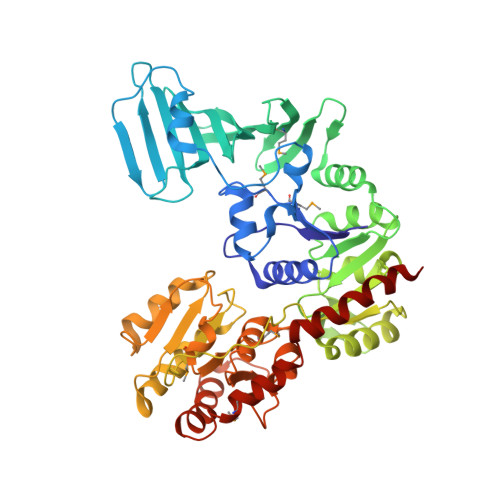
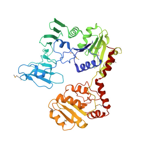
    O-glycosylation of Ser and Thr residues is an important process in all organisms, which is only poorly understood. Such modification is required for the export and function of adhesin proteins that mediate the attachment of pathogenic Gram-positive bacteria to host cells. Here, we have analyzed the mechanism by which the cytosolic O-glycosyltransferase GtfA/B of Streptococcus gordonii modifies the Ser/Thr-rich repeats of adhesin. The enzyme is a tetramer containing two molecules each of GtfA and GtfB. The two subunits have the same fold, but only GtfA contains an active site, whereas GtfB provides the primary binding site for adhesin. During a first phase of glycosylation, the conformation of GtfB is restrained by GtfA to bind substrate with unmodified Ser/Thr residues. In a slow second phase, GtfB recognizes residues that are already modified with N-acetylglucosamine, likely by converting into a relaxed conformation in which one interface with GtfA is broken. These results explain how the glycosyltransferase modifies a progressively changing substrate molecule.


  • Organizational Affiliation

    Howard Hughes Medical Institute, Harvard Medical School, Boston, MA 02115; Department of Cell Biology, Harvard Medical School, Boston, MA 02115;


Macromolecules
Find similar proteins by:  (by identity cutoff)  |  3D Structure
Entity ID: 1
MoleculeChains Sequence LengthOrganismDetailsImage
Glycosyltransferase Gtf1
A, C, E, G
503Streptococcus gordoniiMutation(s): 0 
Gene Names: gtf1gtfA
EC: 2.4.1
UniProt
Find proteins for Q9AET5 (Streptococcus gordonii)
Explore Q9AET5 
Go to UniProtKB:  Q9AET5
Entity Groups  
Sequence Clusters30% Identity50% Identity70% Identity90% Identity95% Identity100% Identity
UniProt GroupQ9AET5
Sequence Annotations
Expand
  • Reference Sequence
Find similar proteins by:  (by identity cutoff)  |  3D Structure
Entity ID: 2
MoleculeChains Sequence LengthOrganismDetailsImage
Glycosyltransferase-stabilizing protein Gtf2
B, D, F, H
446Streptococcus gordoniiMutation(s): 0 
Gene Names: gtf2gtfB
UniProt
Find proteins for Q79T00 (Streptococcus gordonii)
Explore Q79T00 
Go to UniProtKB:  Q79T00
Entity Groups  
Sequence Clusters30% Identity50% Identity70% Identity90% Identity95% Identity100% Identity
UniProt GroupQ79T00
Sequence Annotations
Expand
  • Reference Sequence
Small Molecules
Ligands 3 Unique
IDChains Name / Formula / InChI Key2D Diagram3D Interactions
UDP
Query on UDP

Download Ideal Coordinates CCD File 
I [auth A],
N [auth E]
URIDINE-5'-DIPHOSPHATE
C9 H14 N2 O12 P2
XCCTYIAWTASOJW-XVFCMESISA-N
NAG
Query on NAG

Download Ideal Coordinates CCD File 
J [auth A],
O [auth E]
2-acetamido-2-deoxy-beta-D-glucopyranose
C8 H15 N O6
OVRNDRQMDRJTHS-FMDGEEDCSA-N
MG
Query on MG

Download Ideal Coordinates CCD File 
K [auth B],
L [auth D],
M [auth E],
P [auth G]
MAGNESIUM ION
Mg
JLVVSXFLKOJNIY-UHFFFAOYSA-N
Modified Residues  1 Unique
IDChains TypeFormula2D DiagramParent
MSE
Query on MSE
A, C, E, G
L-PEPTIDE LINKINGC5 H11 N O2 SeMET
Experimental Data & Validation

Experimental Data

  • Method: X-RAY DIFFRACTION
  • Resolution: 3.84 Å
  • R-Value Free: 0.337 
  • R-Value Work: 0.269 
  • R-Value Observed: 0.271 
  • Space Group: P 21 21 21
Unit Cell:
Length ( Å )Angle ( ˚ )
a = 148.975α = 90
b = 196.005β = 90
c = 215.673γ = 90
Software Package:
Software NamePurpose
PHENIXrefinement
Aimlessdata scaling
PDB_EXTRACTdata extraction
XDSdata reduction
PHASERphasing
Cootmodel building

Structure Validation

View Full Validation Report


Ligand Structure Quality Assessment 


Entry History & Funding Information

Deposition Data


Funding OrganizationLocationGrant Number
National Institutes of Health/National Institute of General Medical Sciences (NIH/NIGMS)United StatesGM052586
Howard Hughes Medical Institute (HHMI)United States--

Revision History  (Full details and data files)

  • Version 1.0: 2016-03-02
    Type: Initial release
  • Version 1.1: 2016-03-16
    Changes: Database references
  • Version 1.2: 2017-09-27
    Changes: Author supporting evidence, Database references, Derived calculations
  • Version 1.3: 2019-11-20
    Changes: Author supporting evidence
  • Version 1.4: 2020-07-29
    Type: Remediation
    Reason: Carbohydrate remediation
    Changes: Data collection, Derived calculations, Structure summary
  • Version 1.5: 2023-09-27
    Changes: Data collection, Database references, Refinement description, Structure summary
  • Version 1.6: 2023-11-15
    Changes: Data collection