


On July 21, 2027, wwPDB will fully transition to extended 12-character PDB IDs, the PDBx/mmCIF format, and the re-organized PDB archive.
Start using the PDB Beta Archive today.
RCSB Protein Data Bank (RCSB PDB) enables breakthroughs in science and education by providing access and tools for exploration, visualization, and analysis of:
Experimentally-determined 3D structures from the Protein Data Bank (PDB) archive | |
Integrative 3D Structures from the PDB Archive | |
Computed Structure Models (CSM) from AlphaFold DB and ModelArchive |


Latest EntriesAs ofWed Jul 22 2026

horse myoglobin amyloid fibril - PM2
SpACSA with propyl-AMP and CoA
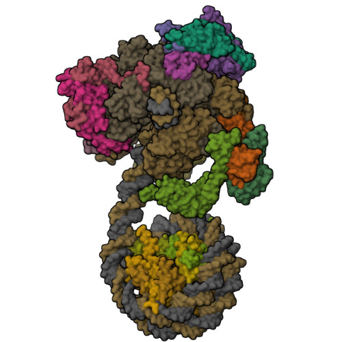
RNA polymerase II elongation complex with the +1 nucleosome

C2 expanded and subtracted 20S Alpha 3 Deletion proteasome core particle in complex with Blm10, Halfmer
Structure of the Ancestral Haloarchaeal (Anc80) Malate dehydrogenase with chloride and potassium ions.
Cryo-EM structure of apo-STING mutant
Crystal Structure of IDH1 (R132H) in complex with covalent inhibitor LY3410738
X-ray structure of the adduct formed upon reaction of the gold compound AF-Npx with lysozyme (Structure 2)

SMAD4 MH2, residues 272-552

Variant T321A of Orotidine 5'-monophosphate decarboxylase-domain of human UMPS in resting state
Features & Highlights

Big PDB Changes Are One Year Away: Extended PDB IDs, the PDB Beta Archive, and PDBx/mmCIF
On July 21, 2027, the wwPDB will fully transition to extended PDB IDs for new depositions and the PDB archive will use a new directory structure based on 12-character extended PDB IDs. Users should adopt these upcoming changes today.

Register for the July 30 Webinar on the AI-Powered 3D Structure Similarity Search
Learn how to use this new feature that delivers structural comparisons across experimentally determined and predicted Computed Structure Models (CSMs).

Register for the December 7 Webinar on pdb_extract
The newly redesigned pdb_extract pre-deposition service streamlines the preparation of structure deposition

Register for the Nov 16 Virtual Office Hour on Visualization
Learn how to visualize molecules large and small at RCSB.org

Register for the Sept 17 Webinar on Remediated Metalloprotein Data in the PDB
Learn how metalloprotein data have been enhanced to support Findability, Accessibility, Interoperability, and Reusability of PDB data

Register for the Nov 3 Ask A Biocurator Anything
Bring your questions about Deposition, Validation, and or Biocuration to this Office Hour. Biocurators will answer general questions about the process or about your specific entry or session.

Register for the Oct 22 Virtual Office Hour on Transitioning to the New PDB Beta Archive, Extended PDB IDs, and PDBx/mmCIF
Learn how to prepare for the July 21, 2027 transition, including using shortlinks to in your code. This Office Hour is for all users, but particularly for software developers and journal editors.

Improved OneDep Support for 3D ED/MicroED
3D ED/MicroED Electron Crystallography metadata are now collected and represented using a macromolecular crystallography data model co-developed by the community.
RCSB.org Help and Documentation
Search or browse the collection of articles designed to help users explore the world of 3D biomolecular structures

Join our DevOps Team at Rutgers
RCSB PDB is looking for an IT Infrastructure Lead to manage our team of systems administrators and DevOps engineers, and support the ongoing operations and growth of our IT infrastructure.
See new feature archive

PDB-101 Focus: Biotechnology
PDB-101 materials explore how researchers are using biology in industry, including how biological evolution is being harnessed in the lab to create new enzymes
» 07/20/2026
Meet RCSB PDB at The Protein Society
Catch up on recent developments in the poster session, and join us in celebrating new Protein Society Fellows Stephen Burley and Helen Berman.
» 07/15/2026Summer Newsletter Published
Highlights Extended PDB IDs; updates to predicted Computer Structure Models at RCSB.org; and PDB-101 resources for exploring protein domains. In the Education Corner, learn about Solving Structures, Enabling Discovery: The Impact of the CRSTAL-ID Centers with SSGCID and CSBID.
» 07/15/2026
Meet the RCSB PDB at ISMB
Join us for posters and presentations at the Conference on Intelligent Systems for Molecular Biology. Interested in joining our team? Talk to us to learn more.
» 07/08/2026Watch the Webinar: Exploring the Workhorses of Biotechnology
Learn how to use RCSB.org to analyze plastic-degrading enzymes
» 06/29/2026
PDB-101 Focus: Biotechnology
PDB-101 materials explore how researchers are using biology in industry. 3D print alpha-amylase, which is used in large quantities in the production of high fructose corn syrup
» 06/22/2026
Watch the Virtual Office Hour on Structure Summary Pages
Learn how a exploring a single PDB structure at RCSB.org can lead to visualizations, analyses, and searches across the archive
» 06/16/2026
Watch the Tutorial: Introduction to Molecular Animation
Learn how to use ChimeraX to create simple animations that can enrich your presentations, websites, and social media
» 06/09/2026
Molecular Origami: Build 3D Paper Models of Protein Domains
Download printable alpha helices and beta strand templates for hands-on exploration of protein folding domains, including a TIM Barrel and a Beta Sandwich
» 06/02/2026
Video: Immunology and Cancer
Visit PDB-101 to watch a three-part series exploring the cellular and molecular details of the human immune response to cancer. Now available in English and Spanish.
» 05/25/2026Molecule of the Month

Quarterly News (see archive)
Issue 110 - July 2026
This issue highlights Extended PDB IDs; updates to predicted Computer Structure Models at RCSB.org, and PDB-101 resources for exploring protein domains.
Annual Reports

2025 Annual Report
Download the 2025 Annual Report (PDF) for an overview of recent activities and the global impact of PDB data and RCSB PDB services.


