8TI1

Cryo-EM structure of a SUR1/Kir6.2-Q52R ATP-sensitive potassium channel in the presence of PIP2 in the open conformation


Experimental Data Snapshot

  • Method: ELECTRON MICROSCOPY
  • Resolution: 2.90 Å
  • Aggregation State: PARTICLE 
  • Reconstruction Method: SINGLE PARTICLE 

wwPDB Validation   3D Report Full Report


This is version 1.0 of the entry. See complete history


Literature

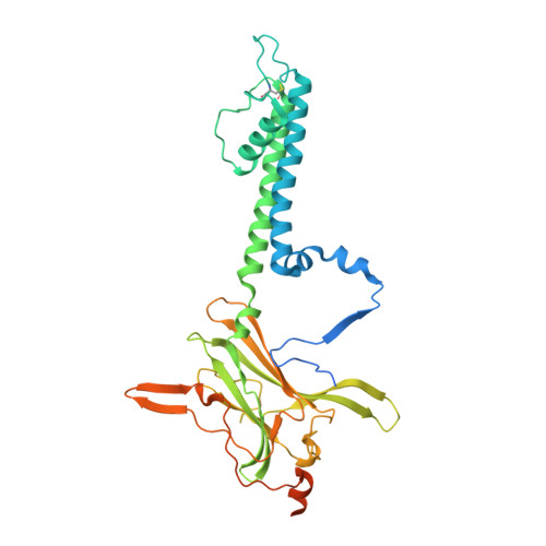
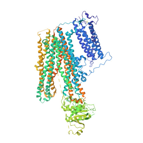
Structure of an open K ATP channel reveals tandem PIP 2 binding sites mediating the Kir6.2 and SUR1 regulatory interface.

Driggers, C.M.Kuo, Y.Y.Zhu, P.ElSheikh, A.Shyng, S.L.

(2024) Nat Commun 15: 2502-2502

  • DOI: https://doi.org/10.1038/s41467-024-46751-5
  • Primary Citation of Related Structures:  
    8TI1, 8TI2

  • PubMed Abstract: 

    ATP-sensitive potassium (K ATP ) channels, composed of four pore-lining Kir6.2 subunits and four regulatory sulfonylurea receptor 1 (SUR1) subunits, control insulin secretion in pancreatic β-cells. K ATP channel opening is stimulated by PIP 2 and inhibited by ATP. Mutations that increase channel opening by PIP 2 reduce ATP inhibition and cause neonatal diabetes. Although considerable evidence has implicated a role for PIP 2 in K ATP channel function, previously solved open-channel structures have lacked bound PIP 2 , and mechanisms by which PIP 2 regulates K ATP channels remain unresolved. Here, we report the cryoEM structure of a K ATP channel harboring the neonatal diabetes mutation Kir6.2-Q52R, in the open conformation, bound to amphipathic molecules consistent with natural C18:0/C20:4 long-chain PI(4,5)P 2 at two adjacent binding sites between SUR1 and Kir6.2. The canonical PIP 2 binding site is conserved among PIP 2 -gated Kir channels. The non-canonical PIP 2 binding site forms at the interface of Kir6.2 and SUR1. Functional studies demonstrate both binding sites determine channel activity. Kir6.2 pore opening is associated with a twist of the Kir6.2 cytoplasmic domain and a rotation of the N-terminal transmembrane domain of SUR1, which widens the inhibitory ATP binding pocket to disfavor ATP binding. The open conformation is particularly stabilized by the Kir6.2-Q52R residue through cation-π bonding with SUR1-W51. Together, these results uncover the cooperation between SUR1 and Kir6.2 in PIP 2 binding and gating, explain the antagonistic regulation of K ATP channels by PIP 2 and ATP, and provide a putative mechanism by which Kir6.2-Q52R stabilizes an open channel to cause neonatal diabetes.


  • Organizational Affiliation

    Department of Chemical Physiology and Biochemistry, School of Medicine, Oregon Health & Science University, Portland, OR, 97239, USA. driggerc@ohsu.edu.


Macromolecules
Find similar proteins by:  (by identity cutoff)  |  3D Structure
Entity ID: 1
MoleculeChains Sequence LengthOrganismDetailsImage
ATP-sensitive inward rectifier potassium channel 11A,
C [auth B],
D [auth C],
E [auth D]
390Rattus norvegicusMutation(s): 1 
Gene Names: Kcnj11
Membrane Entity: Yes 
UniProt
Find proteins for P70673 (Rattus norvegicus)
Explore P70673 
Go to UniProtKB:  P70673
Entity Groups  
Sequence Clusters30% Identity50% Identity70% Identity90% Identity95% Identity100% Identity
UniProt GroupP70673
Sequence Annotations
Expand
  • Reference Sequence
Find similar proteins by:  (by identity cutoff)  |  3D Structure
Entity ID: 2
MoleculeChains Sequence LengthOrganismDetailsImage
SUR1B [auth E],
F [auth H],
G,
H [auth F]
1,582Mesocricetus auratusMutation(s): 0 
Membrane Entity: Yes 
UniProt
Find proteins for A0A1S4NYG1 (Mesocricetus auratus)
Explore A0A1S4NYG1 
Go to UniProtKB:  A0A1S4NYG1
Entity Groups  
Sequence Clusters30% Identity50% Identity70% Identity90% Identity95% Identity100% Identity
UniProt GroupA0A1S4NYG1
Sequence Annotations
Expand
  • Reference Sequence
Small Molecules
Ligands 5 Unique
IDChains Name / Formula / InChI Key2D Diagram3D Interactions
PT5 (Subject of Investigation/LOI)
Query on PT5

Download Ideal Coordinates CCD File 
AA [auth G]
FA [auth F]
I [auth A]
N [auth E]
S [auth B]
AA [auth G],
FA [auth F],
I [auth A],
N [auth E],
S [auth B],
T [auth C],
U [auth D],
V [auth H]
[(2R)-1-octadecanoyloxy-3-[oxidanyl-[(1R,2R,3S,4R,5R,6S)-2,3,6-tris(oxidanyl)-4,5-diphosphonooxy-cyclohexyl]oxy-phospho ryl]oxy-propan-2-yl] (8Z)-icosa-5,8,11,14-tetraenoate
C47 H85 O19 P3
CNWINRVXAYPOMW-HJBQCNPJSA-N
P5S
Query on P5S

Download Ideal Coordinates CCD File 
CA [auth G]
DA [auth G]
HA [auth F]
IA [auth F]
P [auth E]
CA [auth G],
DA [auth G],
HA [auth F],
IA [auth F],
P [auth E],
Q [auth E],
X [auth H],
Y [auth H]
O-[(R)-{[(2R)-2,3-bis(octadecanoyloxy)propyl]oxy}(hydroxy)phosphoryl]-L-serine
C42 H82 N O10 P
TZCPCKNHXULUIY-RGULYWFUSA-N
PEF
Query on PEF

Download Ideal Coordinates CCD File 
BA [auth G],
GA [auth F],
O [auth E],
W [auth H]
DI-PALMITOYL-3-SN-PHOSPHATIDYLETHANOLAMINE
C37 H74 N O8 P
SLKDGVPOSSLUAI-PGUFJCEWSA-N
NAG
Query on NAG

Download Ideal Coordinates CCD File 
EA [auth G],
JA [auth F],
R [auth E],
Z [auth H]
2-acetamido-2-deoxy-beta-D-glucopyranose
C8 H15 N O6
OVRNDRQMDRJTHS-FMDGEEDCSA-N
K
Query on K

Download Ideal Coordinates CCD File 
J [auth A],
K [auth A],
L [auth A],
M [auth A]
POTASSIUM ION
K
NPYPAHLBTDXSSS-UHFFFAOYSA-N
Experimental Data & Validation

Experimental Data

  • Method: ELECTRON MICROSCOPY
  • Resolution: 2.90 Å
  • Aggregation State: PARTICLE 
  • Reconstruction Method: SINGLE PARTICLE 
EM Software:
TaskSoftware PackageVersion
MODEL REFINEMENTPHENIX
RECONSTRUCTIONcryoSPARC4.2.1

Structure Validation

View Full Validation Report


Entry History & Funding Information

Deposition Data


Funding OrganizationLocationGrant Number
National Institutes of Health/National Institute of Diabetes and Digestive and Kidney Disease (NIH/NIDDK)United StatesDK066485
National Institutes of Health/National Institute of General Medical Sciences (NIH/NIGMS)United StatesGM129547

Revision History  (Full details and data files)

  • Version 1.0: 2024-04-03
    Type: Initial release