8J1Q | pdb_00008j1q

CryoEM structure of SARS CoV-2 RBD and Aptamer complex


Experimental Data Snapshot

  • Method: ELECTRON MICROSCOPY
  • Resolution: 3.30 Å
  • Aggregation State: PARTICLE 
  • Reconstruction Method: SINGLE PARTICLE 

Starting Models: experimental
View more details

wwPDB Validation 3D Report Full Report

Validation slider image for 8J1Q

This is version 1.2 of the entry. See complete history

Literature

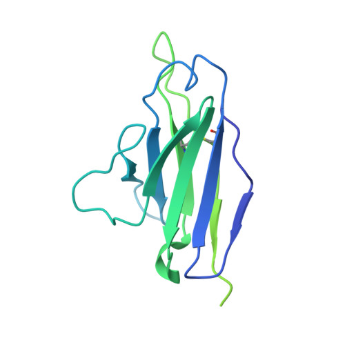
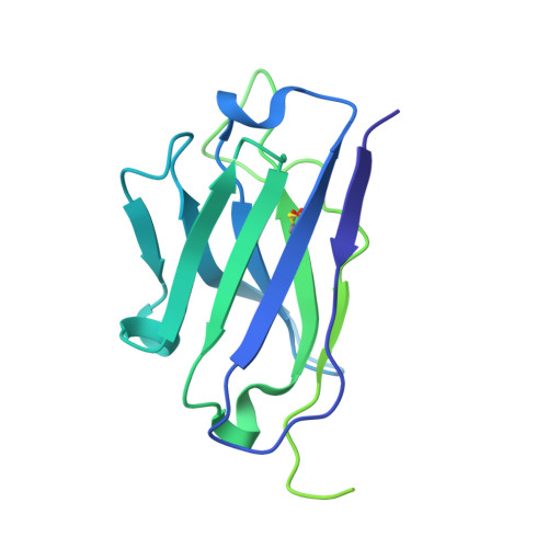
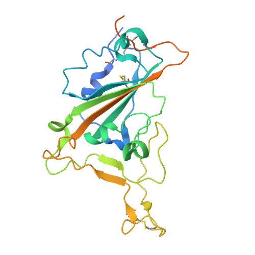
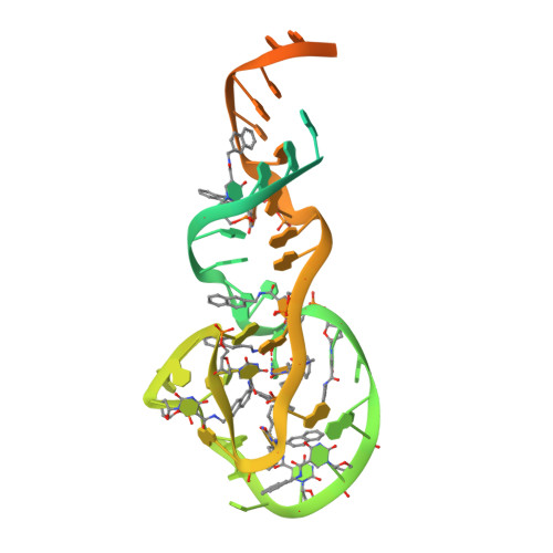
Structure-Guided Development of Bivalent Aptamers Blocking SARS-CoV-2 Infection.

Rahman, M.S.Han, M.J.Kim, S.W.Kang, S.M.Kim, B.R.Kim, H.Lee, C.J.Noh, J.E.Kim, H.Lee, J.O.Jang, S.K.

(2023) Molecules 28

  • DOI: https://doi.org/10.3390/molecules28124645
  • Primary Citation Related Structures: 
    8J1Q, 8J26

  • PubMed Abstract: 

    Severe acute respiratory syndrome coronavirus 2 (SARS-CoV-2) has caused devastation to human society through its high virulence, infectivity, and genomic mutations, which reduced the efficacy of vaccines. Here, we report the development of aptamers that effectively interfere with SARS-CoV-2 infection by targeting its spike protein, which plays a pivotal role in host cell entry of the virus through interaction with the viral receptor angiotensin-converting enzyme 2 (ACE2). To develop highly effective aptamers and to understand their mechanism in inhibiting viral infection, we determined the three-dimensional (3D) structures of aptamer/receptor-binding domain (RBD) complexes using cryogenic electron microscopy (cryo-EM). Moreover, we developed bivalent aptamers targeting two distinct regions of the RBD in the spike protein that directly interact with ACE2. One aptamer interferes with the binding of ACE2 by blocking the ACE2-binding site in RBD, and the other aptamer allosterically inhibits ACE2 by binding to a distinct face of RBD. Using the 3D structures of aptamer-RBD complexes, we minimized and optimized these aptamers. By combining the optimized aptamers, we developed a bivalent aptamer that showed a stronger inhibitory effect on virus infection than the component aptamers. This study confirms that the structure-based aptamer-design approach has a high potential in developing antiviral drugs against SARS-CoV-2 and other viruses.


  • Organizational Affiliation
    • Department of Life Sciences, POSTECH Biotech Center, Pohang University of Science and Technology, 77 Cheongam-ro, Nam-gu, Pohang-si 37673, Republic of Korea.

Macromolecule Content 

  • Total Structure Weight: 123.8 kDa 
  • Atom Count: 5,447 
  • Modeled Residue Count: 517 
  • Deposited Residue Count: 857 
  • Unique protein chains: 3
  • Unique nucleic acid chains: 2

Macromolecules


Find similar proteins by:|  3D Structure
Entity ID: 3
MoleculeChains  Sequence LengthOrganismDetailsImage
Fab Light chain (REGN10987)C [auth A]224Homo sapiensMutation(s): 0 
Find similar proteins by:|  3D Structure
Entity ID: 4
MoleculeChains  Sequence LengthOrganismDetailsImage
Fab heavy chain (REGN10987)D [auth B]248Homo sapiensMutation(s): 0 
Find similar proteins by:|  3D Structure
Entity ID: 5
MoleculeChains  Sequence LengthOrganismDetailsImage
Spike protein S1E [auth C]253Severe acute respiratory syndrome coronavirus 2Mutation(s): 0 
Gene Names: S2
UniProt
Find proteins for P0DTC2 (Severe acute respiratory syndrome coronavirus 2)
Explore P0DTC2 
Go to UniProtKB:  P0DTC2
Entity Groups
Sequence Clusters30% Identity50% Identity70% Identity90% Identity95% Identity100% Identity
UniProt GroupP0DTC2
Glycosylation
Glycosylation Sites: 1Go to GlyGen: P0DTC2-1
Sequence Annotations
Expand
Reference Sequence
Find similar nucleic acids by:  (by identity cutoff) 
Entity ID: 1
MoleculeChains LengthOrganismImage
AM032-0A [auth E]80synthetic construct
Sequence Annotations
Expand
Reference Sequence
Find similar nucleic acids by:  (by identity cutoff) 
Entity ID: 2
MoleculeChains LengthOrganismImage
AM047-0B [auth D]52synthetic construct
Sequence Annotations
Expand
Reference Sequence

Small Molecules

Ligands 1 Unique
IDChains Name / Formula / InChI Key2D Diagram3D Interactions
NAG
(Subject of Investigation/LOI)

Query on NAG


Download:Ideal Coordinates CCD File
F [auth C]2-acetamido-2-deoxy-beta-D-glucopyranose
C8 H15 N O6
OVRNDRQMDRJTHS-FMDGEEDCSA-N

Experimental Data & Validation

Experimental Data

  • Method: ELECTRON MICROSCOPY
  • Resolution: 3.30 Å
  • Aggregation State: PARTICLE 
  • Reconstruction Method: SINGLE PARTICLE 
EM Software:
TaskSoftware PackageVersion
MODEL REFINEMENTPHENIX1.21rc1_4895:
MODEL REFINEMENTCoot0.9.6 EL
RECONSTRUCTIONcryoSPARC4.1.0 and 4.2.1

Structure Validation

View Full Validation Report


Entry History 

& Funding Information

Deposition Data


Funding OrganizationLocationGrant Number
National Research Foundation (NRF, Korea)Korea, Republic OfNRF-2019M3E5D6063871

Revision History  (Full details and data files)

  • Version 1.0: 2023-06-21
    Type: Initial release
  • Version 1.1: 2023-07-12
    Changes: Database references
  • Version 1.2: 2024-11-20
    Changes: Data collection, Refinement description, Structure summary