8IJX

Cryo-EM structure of the gastric proton pump with bound DQ-18


Experimental Data Snapshot

  • Method: ELECTRON MICROSCOPY
  • Resolution: 2.08 Å
  • Aggregation State: PARTICLE 
  • Reconstruction Method: SINGLE PARTICLE 

wwPDB Validation   3D Report Full Report


This is version 1.1 of the entry. See complete history


Literature

Deep learning driven de novo drug design based on gastric proton pump structures.

Abe, K.Ozako, M.Inukai, M.Matsuyuki, Y.Kitayama, S.Kanai, C.Nagai, C.Gopalasingam, C.C.Gerle, C.Shigematsu, H.Umekubo, N.Yokoshima, S.Yoshimori, A.

(2023) Commun Biol 6: 956-956

  • DOI: https://doi.org/10.1038/s42003-023-05334-8
  • Primary Citation of Related Structures:  
    8IJV, 8IJW, 8IJX, 8JMN

  • PubMed Abstract: 

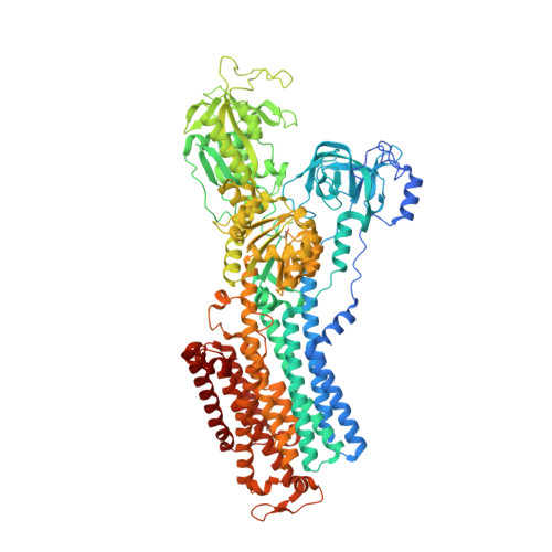
    Existing drugs often suffer in their effectiveness due to detrimental side effects, low binding affinity or pharmacokinetic problems. This may be overcome by the development of distinct compounds. Here, we exploit the rich structural basis of drug-bound gastric proton pump to develop compounds with strong inhibitory potency, employing a combinatorial approach utilizing deep generative models for de novo drug design with organic synthesis and cryo-EM structural analysis. Candidate compounds that satisfy pharmacophores defined in the drug-bound proton pump structures, were designed in silico utilizing our deep generative models, a workflow termed Deep Quartet. Several candidates were synthesized and screened according to their inhibition potencies in vitro, and their binding poses were in turn identified by cryo-EM. Structures reaching up to 2.10 Å resolution allowed us to evaluate and re-design compound structures, heralding the most potent compound in this study, DQ-18 (N-methyl-4-((2-(benzyloxy)-5-chlorobenzyl)oxy)benzylamine), which shows a K i value of 47.6 nM. Further high-resolution cryo-EM analysis at 2.08 Å resolution unambiguously determined the DQ-18 binding pose. Our integrated approach offers a framework for structure-based de novo drug development based on the desired pharmacophores within the protein structure.


  • Organizational Affiliation

    Cellular and Structural Physiology Institute, Nagoya University, Nagoya, Aichi, 464-8601, Japan. kabe@cespi.nagoya-u.ac.jp.


Macromolecules
Find similar proteins by:  (by identity cutoff)  |  3D Structure
Entity ID: 1
MoleculeChains Sequence LengthOrganismDetailsImage
Sodium/potassium-transporting ATPase subunit alpha1,033Sus scrofaMutation(s): 1 
Gene Names: ATP4A
Membrane Entity: Yes 
UniProt
Find proteins for P19156 (Sus scrofa)
Explore P19156 
Go to UniProtKB:  P19156
Entity Groups  
Sequence Clusters30% Identity50% Identity70% Identity90% Identity95% Identity100% Identity
UniProt GroupP19156
Sequence Annotations
Expand
  • Reference Sequence
Find similar proteins by:  (by identity cutoff)  |  3D Structure
Entity ID: 2
MoleculeChains Sequence LengthOrganismDetailsImage
Potassium-transporting ATPase subunit beta290Sus scrofaMutation(s): 0 
Gene Names: ATP4B
Membrane Entity: Yes 
UniProt
Find proteins for P18434 (Sus scrofa)
Explore P18434 
Go to UniProtKB:  P18434
Entity Groups  
Sequence Clusters30% Identity50% Identity70% Identity90% Identity95% Identity100% Identity
UniProt GroupP18434
Sequence Annotations
Expand
  • Reference Sequence
Small Molecules
Ligands 5 Unique
IDChains Name / Formula / InChI Key2D Diagram3D Interactions
PCW
Query on PCW

Download Ideal Coordinates CCD File 
C [auth A]
E [auth A]
F [auth A]
G [auth A]
I [auth A]
C [auth A],
E [auth A],
F [auth A],
G [auth A],
I [auth A],
J [auth B]
1,2-DIOLEOYL-SN-GLYCERO-3-PHOSPHOCHOLINE
C44 H85 N O8 P
SNKAWJBJQDLSFF-NVKMUCNASA-O
CLR
Query on CLR

Download Ideal Coordinates CCD File 
N [auth B]CHOLESTEROL
C27 H46 O
HVYWMOMLDIMFJA-DPAQBDIFSA-N
PZ0
Query on PZ0

Download Ideal Coordinates CCD File 
H [auth A]1-[4-[(5-chloranyl-2-phenylmethoxy-phenyl)methoxy]phenyl]-N-methyl-methanamine
C22 H22 Cl N O2
KKMPHNRWOFIHIE-UHFFFAOYSA-N
NAG
Query on NAG

Download Ideal Coordinates CCD File 
K [auth B],
L [auth B],
M [auth B]
2-acetamido-2-deoxy-beta-D-glucopyranose
C8 H15 N O6
OVRNDRQMDRJTHS-FMDGEEDCSA-N
MG
Query on MG

Download Ideal Coordinates CCD File 
D [auth A]MAGNESIUM ION
Mg
JLVVSXFLKOJNIY-UHFFFAOYSA-N
Modified Residues  1 Unique
IDChains TypeFormula2D DiagramParent
BFD
Query on BFD
A
L-PEPTIDE LINKINGC4 H6 Be F3 N O4ASP
Experimental Data & Validation

Experimental Data

  • Method: ELECTRON MICROSCOPY
  • Resolution: 2.08 Å
  • Aggregation State: PARTICLE 
  • Reconstruction Method: SINGLE PARTICLE 

Structure Validation

View Full Validation Report


Entry History & Funding Information

Deposition Data


Funding OrganizationLocationGrant Number
Japan Society for the Promotion of Science (JSPS)Japan21H02426

Revision History  (Full details and data files)

  • Version 1.0: 2023-08-30
    Type: Initial release
  • Version 1.1: 2023-10-04
    Changes: Database references