8EGX

Complex of Fat4(EC1-4) bound to Dchs1(EC1-4)

  • Classification: CELL ADHESION
  • Organism(s): Homo sapiens
  • Expression System: Trichoplusia ni
  • Mutation(s): No 

  • Deposited: 2022-09-13 Released: 2023-02-22 
  • Deposition Author(s): Medina, E., Luca, V.C.
  • Funding Organization(s): National Institutes of Health/National Institute of General Medical Sciences (NIH/NIGMS)

Experimental Data Snapshot

  • Method: X-RAY DIFFRACTION
  • Resolution: 3.69 Å
  • R-Value Free: 0.298 
  • R-Value Work: 0.271 
  • R-Value Observed: 0.272 

wwPDB Validation   3D Report Full Report


Ligand Structure Quality Assessment 


This is version 1.2 of the entry. See complete history


Literature

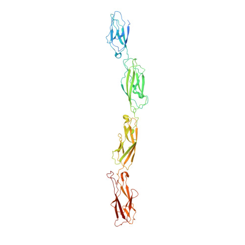
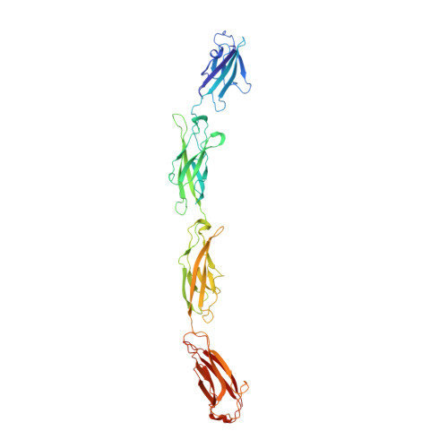
Structure of the planar cell polarity cadherins Fat4 and Dachsous1.

Medina, E.Easa, Y.Lester, D.K.Lau, E.K.Sprinzak, D.Luca, V.C.

(2023) Nat Commun 14: 891-891

  • DOI: https://doi.org/10.1038/s41467-023-36435-x
  • Primary Citation of Related Structures:  
    8EGW, 8EGX

  • PubMed Abstract: 

    The atypical cadherins Fat and Dachsous are key regulators of cell growth and animal development. In contrast to classical cadherins, which form homophilic interactions to segregate cells, Fat and Dachsous cadherins form heterophilic interactions to induce cell polarity within tissues. Here, we determine the co-crystal structure of the human homologs Fat4 and Dachsous1 (Dchs1) to establish the molecular basis for Fat-Dachsous interactions. The binding domains of Fat4 and Dchs1 form an extended interface along extracellular cadherin (EC) domains 1-4 of each protein. Biophysical measurements indicate that Fat4-Dchs1 affinity is among the highest reported for cadherin superfamily members, which is attributed to an extensive network of salt bridges not present in structurally similar protocadherin homodimers. Furthermore, modeling suggests that unusual extracellular phosphorylation modifications directly modulate Fat-Dachsous binding by introducing charged contacts across the interface. Collectively, our analyses reveal how the molecular architecture of Fat4-Dchs1 enables them to form long-range, high-affinity interactions to maintain planar cell polarity.


  • Organizational Affiliation

    Department of Drug Discovery, H. Lee Moffitt Cancer Center & Research Institute, Tampa, FL, 33602, USA.


Macromolecules
Find similar proteins by:  (by identity cutoff)  |  3D Structure
Entity ID: 1
MoleculeChains Sequence LengthOrganismDetailsImage
Protocadherin Fat 4A [auth B]467Homo sapiensMutation(s): 0 
Gene Names: FAT4CDHF14FATJNbla00548
UniProt & NIH Common Fund Data Resources
Find proteins for Q6V0I7 (Homo sapiens)
Explore Q6V0I7 
Go to UniProtKB:  Q6V0I7
PHAROS:  Q6V0I7
GTEx:  ENSG00000196159 
Entity Groups  
Sequence Clusters30% Identity50% Identity70% Identity90% Identity95% Identity100% Identity
UniProt GroupQ6V0I7
Sequence Annotations
Expand
  • Reference Sequence
Find similar proteins by:  (by identity cutoff)  |  3D Structure
Entity ID: 2
MoleculeChains Sequence LengthOrganismDetailsImage
Protocadherin-16B [auth A]421Homo sapiensMutation(s): 0 
Gene Names: DCHS1CDH19CDH25FIB1KIAA1773PCDH16
UniProt & NIH Common Fund Data Resources
Find proteins for Q96JQ0 (Homo sapiens)
Explore Q96JQ0 
Go to UniProtKB:  Q96JQ0
PHAROS:  Q96JQ0
GTEx:  ENSG00000166341 
Entity Groups  
Sequence Clusters30% Identity50% Identity70% Identity90% Identity95% Identity100% Identity
UniProt GroupQ96JQ0
Sequence Annotations
Expand
  • Reference Sequence
Oligosaccharides

Help

Entity ID: 3
MoleculeChains Length2D Diagram Glycosylation3D Interactions
2-acetamido-2-deoxy-beta-D-glucopyranose-(1-4)-2-acetamido-2-deoxy-beta-D-glucopyranose
C
2N-Glycosylation
Glycosylation Resources
GlyTouCan:  G42666HT
GlyCosmos:  G42666HT
GlyGen:  G42666HT
Small Molecules
Ligands 2 Unique
IDChains Name / Formula / InChI Key2D Diagram3D Interactions
NAG
Query on NAG

Download Ideal Coordinates CCD File 
D [auth B],
E [auth B],
O [auth A],
P [auth A]
2-acetamido-2-deoxy-beta-D-glucopyranose
C8 H15 N O6
OVRNDRQMDRJTHS-FMDGEEDCSA-N
CA
Query on CA

Download Ideal Coordinates CCD File 
F [auth B]
G [auth B]
H [auth B]
I [auth B]
J [auth B]
F [auth B],
G [auth B],
H [auth B],
I [auth B],
J [auth B],
K [auth B],
L [auth B],
M [auth B],
N [auth B],
Q [auth A],
R [auth A],
S [auth A],
T [auth A],
U [auth A],
V [auth A],
W [auth A],
X [auth A],
Y [auth A]
CALCIUM ION
Ca
BHPQYMZQTOCNFJ-UHFFFAOYSA-N
Experimental Data & Validation

Experimental Data

  • Method: X-RAY DIFFRACTION
  • Resolution: 3.69 Å
  • R-Value Free: 0.298 
  • R-Value Work: 0.271 
  • R-Value Observed: 0.272 
  • Space Group: P 21 21 21
Unit Cell:
Length ( Å )Angle ( ˚ )
a = 64.222α = 90
b = 82.102β = 90
c = 224.759γ = 90
Software Package:
Software NamePurpose
PHENIXrefinement
PDB_EXTRACTdata extraction
HKL-2000data reduction
HKL-2000data scaling
PHASERphasing

Structure Validation

View Full Validation Report


Ligand Structure Quality Assessment 


Entry History & Funding Information

Deposition Data


Funding OrganizationLocationGrant Number
National Institutes of Health/National Institute of General Medical Sciences (NIH/NIGMS)United StatesR35GM133482

Revision History  (Full details and data files)

  • Version 1.0: 2023-02-22
    Type: Initial release
  • Version 1.1: 2023-03-01
    Changes: Database references
  • Version 1.2: 2023-10-25
    Changes: Data collection, Refinement description