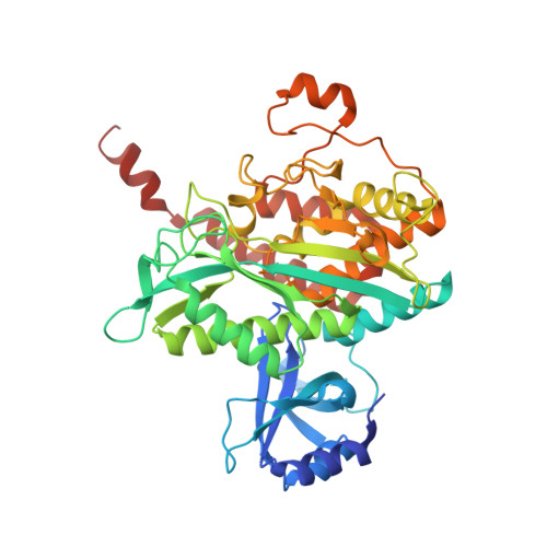
7TDV

Crystal structure of S. aureus glutamine synthetase in Met-Sox-P/ADP transition state complex

  • Classification: LIGASE
  • Organism(s): Staphylococcus aureus
  • Expression System: Escherichia coli
  • Mutation(s): No 

  • Deposited: 2022-01-03 Released: 2022-06-29 
  • Deposition Author(s): Schumacher, M.A.
  • Funding Organization(s): National Institutes of Health/National Institute of General Medical Sciences (NIH/NIGMS)

Experimental Data Snapshot

  • Method: X-RAY DIFFRACTION
  • Resolution: 2.92 Å
  • R-Value Free: 0.256 
  • R-Value Work: 0.198 
  • R-Value Observed: 0.200 

wwPDB Validation   3D Report Full Report


Ligand Structure Quality Assessment 


This is version 1.2 of the entry. See complete history


Literature

Molecular dissection of the glutamine synthetase-GlnR nitrogen regulatory circuitry in Gram-positive bacteria.

Travis, B.A.Peck, J.V.Salinas, R.Dopkins, B.Lent, N.Nguyen, V.D.Borgnia, M.J.Brennan, R.G.Schumacher, M.A.

(2022) Nat Commun 13: 3793-3793

  • DOI: https://doi.org/10.1038/s41467-022-31573-0
  • Primary Citation of Related Structures:  
    7TDP, 7TDV, 7TEA, 7TEC, 7TEN, 7TF6, 7TF7, 7TF9, 7TFA, 7TFB, 7TFC, 7TFD, 7TFE

  • PubMed Abstract: 

    How bacteria sense and respond to nitrogen levels are central questions in microbial physiology. In Gram-positive bacteria, nitrogen homeostasis is controlled by an operon encoding glutamine synthetase (GS), a dodecameric machine that assimilates ammonium into glutamine, and the GlnR repressor. GlnR detects nitrogen excess indirectly by binding glutamine-feedback-inhibited-GS (FBI-GS), which activates its transcription-repression function. The molecular mechanisms behind this regulatory circuitry, however, are unknown. Here we describe biochemical and structural analyses of GS and FBI-GS-GlnR complexes from pathogenic and non-pathogenic Gram-positive bacteria. The structures show FBI-GS binds the GlnR C-terminal domain within its active-site cavity, juxtaposing two GlnR monomers to form a DNA-binding-competent GlnR dimer. The FBI-GS-GlnR interaction stabilizes the inactive GS conformation. Strikingly, this interaction also favors a remarkable dodecamer to tetradecamer transition in some GS, breaking the paradigm that all bacterial GS are dodecamers. These data thus unveil unique structural mechanisms of transcription and enzymatic regulation.


  • Organizational Affiliation

    Department of Biochemistry, 307 Research Dr., Box 3711, Duke University Medical Center, Durham, NC, 27710, USA.


Macromolecules
Find similar proteins by:  (by identity cutoff)  |  3D Structure
Entity ID: 1
MoleculeChains Sequence LengthOrganismDetailsImage
Glutamine synthetase449Staphylococcus aureusMutation(s): 0 
Gene Names: glnABSG37_06900FAF32_003410G6X35_05920G6X37_15495G6Y24_07770SAMEA103891454_00902
EC: 6.3.1.2
UniProt
Find proteins for E3VXC2 (Staphylococcus aureus)
Explore E3VXC2 
Go to UniProtKB:  E3VXC2
Entity Groups  
Sequence Clusters30% Identity50% Identity70% Identity90% Identity95% Identity100% Identity
UniProt GroupE3VXC2
Sequence Annotations
Expand
  • Reference Sequence
Small Molecules
Ligands 4 Unique
IDChains Name / Formula / InChI Key2D Diagram3D Interactions
ADP
Query on ADP

Download Ideal Coordinates CCD File 
CA [auth B]
G [auth A]
JA [auth H]
K [auth C]
R [auth D]
CA [auth B],
G [auth A],
JA [auth H],
K [auth C],
R [auth D],
W [auth E]
ADENOSINE-5'-DIPHOSPHATE
C10 H15 N5 O10 P2
XTWYTFMLZFPYCI-KQYNXXCUSA-N
P3S (Subject of Investigation/LOI)
Query on P3S

Download Ideal Coordinates CCD File 
DA [auth B]
H [auth A]
KA [auth H]
L [auth C]
S [auth D]
DA [auth B],
H [auth A],
KA [auth H],
L [auth C],
S [auth D],
X [auth E]
L-METHIONINE-S-SULFOXIMINE PHOSPHATE
C5 H13 N2 O6 P S
QQFOFBSCSWFFPB-NMAPHRJESA-N
SO4
Query on SO4

Download Ideal Coordinates CCD File 
NA [auth H]SULFATE ION
O4 S
QAOWNCQODCNURD-UHFFFAOYSA-L
MG
Query on MG

Download Ideal Coordinates CCD File 
AA [auth E]
BA [auth E]
EA [auth B]
FA [auth B]
GA [auth B]
AA [auth E],
BA [auth E],
EA [auth B],
FA [auth B],
GA [auth B],
HA [auth B],
I [auth A],
IA [auth B],
J [auth A],
LA [auth H],
M [auth C],
MA [auth H],
N [auth C],
O [auth C],
P [auth C],
Q [auth C],
T [auth D],
U [auth D],
V [auth D],
Y [auth E],
Z [auth E]
MAGNESIUM ION
Mg
JLVVSXFLKOJNIY-UHFFFAOYSA-N
Experimental Data & Validation

Experimental Data

  • Method: X-RAY DIFFRACTION
  • Resolution: 2.92 Å
  • R-Value Free: 0.256 
  • R-Value Work: 0.198 
  • R-Value Observed: 0.200 
  • Space Group: P 41 21 2
Unit Cell:
Length ( Å )Angle ( ˚ )
a = 154.615α = 90
b = 154.615β = 90
c = 299.295γ = 90
Software Package:
Software NamePurpose
XDSdata reduction
SCALAdata scaling
PHASERphasing
PHENIXrefinement
PDB_EXTRACTdata extraction

Structure Validation

View Full Validation Report


Ligand Structure Quality Assessment 


Entry History & Funding Information

Deposition Data


Funding OrganizationLocationGrant Number
National Institutes of Health/National Institute of General Medical Sciences (NIH/NIGMS)United StatesR35GM130290

Revision History  (Full details and data files)

  • Version 1.0: 2022-06-29
    Type: Initial release
  • Version 1.1: 2022-07-13
    Changes: Database references
  • Version 1.2: 2023-10-18
    Changes: Data collection, Refinement description