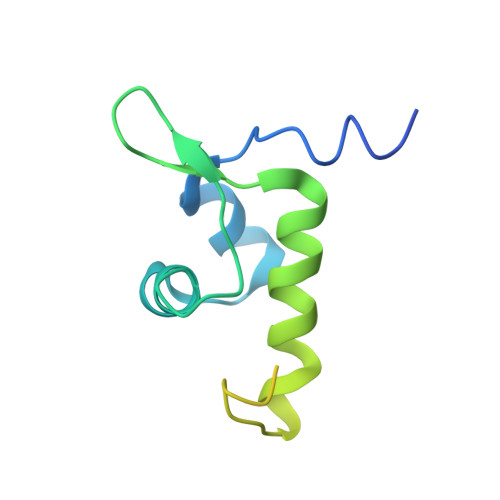
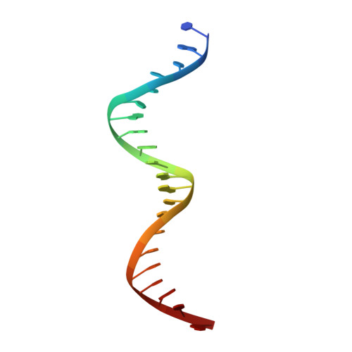
7TEC

Structure of the Listeria monocytogenes GlnR-DNA complex to 3.45 Angstrom


Experimental Data Snapshot

  • Method: X-RAY DIFFRACTION
  • Resolution: 3.45 Å
  • R-Value Free: 0.309 
  • R-Value Work: 0.266 
  • R-Value Observed: 0.270 

wwPDB Validation   3D Report Full Report


This is version 1.2 of the entry. See complete history


Literature

Molecular dissection of the glutamine synthetase-GlnR nitrogen regulatory circuitry in Gram-positive bacteria.

Travis, B.A.Peck, J.V.Salinas, R.Dopkins, B.Lent, N.Nguyen, V.D.Borgnia, M.J.Brennan, R.G.Schumacher, M.A.

(2022) Nat Commun 13: 3793-3793

  • DOI: https://doi.org/10.1038/s41467-022-31573-0
  • Primary Citation of Related Structures:  
    7TDP, 7TDV, 7TEA, 7TEC, 7TEN, 7TF6, 7TF7, 7TF9, 7TFA, 7TFB, 7TFC, 7TFD, 7TFE

  • PubMed Abstract: 

    How bacteria sense and respond to nitrogen levels are central questions in microbial physiology. In Gram-positive bacteria, nitrogen homeostasis is controlled by an operon encoding glutamine synthetase (GS), a dodecameric machine that assimilates ammonium into glutamine, and the GlnR repressor. GlnR detects nitrogen excess indirectly by binding glutamine-feedback-inhibited-GS (FBI-GS), which activates its transcription-repression function. The molecular mechanisms behind this regulatory circuitry, however, are unknown. Here we describe biochemical and structural analyses of GS and FBI-GS-GlnR complexes from pathogenic and non-pathogenic Gram-positive bacteria. The structures show FBI-GS binds the GlnR C-terminal domain within its active-site cavity, juxtaposing two GlnR monomers to form a DNA-binding-competent GlnR dimer. The FBI-GS-GlnR interaction stabilizes the inactive GS conformation. Strikingly, this interaction also favors a remarkable dodecamer to tetradecamer transition in some GS, breaking the paradigm that all bacterial GS are dodecamers. These data thus unveil unique structural mechanisms of transcription and enzymatic regulation.


  • Organizational Affiliation

    Department of Biochemistry, 307 Research Dr., Box 3711, Duke University Medical Center, Durham, NC, 27710, USA.


Macromolecules

Find similar proteins by:  (by identity cutoff)  |  3D Structure
Entity ID: 1
MoleculeChains Sequence LengthOrganismDetailsImage
HTH-type transcriptional regulator GlnR125Listeria monocytogenesMutation(s): 0 
Gene Names: 
glnRA8L61_07010ABZ57_05085AF006_07180AF817_01990AMC55_04605APD94_02400ART25_07755ARV28_05490ARV77_10415B1N06_02425B1N38_06025B1N40_09865B1N52_08095B1N70_08585B1N71_01820B1O10_02055B1O25_08310B2H14_10505B4X79_13085B4Y29_01290B4Y40_11845B4Y47_05380B4Y49_02050B4Y56_02055B4Y57_02055B5K54_13065B6112_07615B6O07_10740BB997_08390BCZ19_06080BW273_06570C6S26_02850CW834_10150CW845_07630CW895_11495CX098_14230D4164_01995D4271_02955D4900_04675D4920_02385D4947_02360D4C60_03330D4D22_02955D4D89_02780D4U00_01965D4U23_10015D5M63_02050D5N24_04355D6P18_02820D7104_08235DCK28_08790DCT16_08455E0I39_02830E1V33_00335E1W43_05380E1W56_05615E3W32_09810E5F58_10230E5H26_02070EPC87_01440EX365_07695EXZ73_06750EYY39_05995F3O35_05110F3R75_05795F6436_09515F6515_11560FA835_14895FC284_14445FJU19_02820FL871_07620FLQ97_14195FLR03_05055FLR11_13675FNX31_12240FNX40_10035FORC68_1320FR217_04555FV747_02470G3O21_002452G3R95_000630GEK29_02375GFK29_08620GH165_00575GHH22_03975GHM39_05980GIG92_01790GIH49_01260GJW51_07325GNG71_06855GON91_01810GT011_02015GXB45_00580GYN46_14485GYO86_08555GYP27_02015GYS19_13395GYU05_01010GYU24_02035GYX95_04495GYY14_11060GYZ23_12950GYZ33_11570GYZ61_07200GZI09_15320GZK40_07615GZM52_07550GZN68_09475HP506_001182HQN34_002361I6I37_04555I8J47_00192IP987_001477JKV73_01561JKV74_07850JKV76_01310JKV77_01646JKX60_08595JKX61_06825KV70_04590KW30_04605LmNIHS28_00654M643_00970QD52_08715R019_12835UI29_08755UP23_04390

UniProt
Find proteins for L8DSZ4 (Listeria monocytogenes)
Explore L8DSZ4 
Go to UniProtKB:  L8DSZ4
Entity Groups  
Sequence Clusters30% Identity50% Identity70% Identity90% Identity95% Identity100% Identity
UniProt GroupL8DSZ4
Sequence Annotations
Expand
  • Reference Sequence

Find similar nucleic acids by:  Sequence   |   3D Structure  

Entity ID: 2
MoleculeChains LengthOrganismImage
DNA (5'-D(*CP*GP*TP*GP*TP*CP*AP*GP*AP*TP*AP*AP*TP*CP*TP*GP*AP*CP*AP*CP*G)-3')B [auth H]21synthetic construct
Sequence Annotations
Expand
  • Reference Sequence
Experimental Data & Validation

Experimental Data

  • Method: X-RAY DIFFRACTION
  • Resolution: 3.45 Å
  • R-Value Free: 0.309 
  • R-Value Work: 0.266 
  • R-Value Observed: 0.270 
  • Space Group: P 2 2 21
Unit Cell:
Length ( Å )Angle ( ˚ )
a = 28.632α = 90
b = 47.717β = 90
c = 121.41γ = 90
Software Package:
Software NamePurpose
PHENIXrefinement
XDSdata reduction
SCALAdata scaling
PHASERphasing
PDB_EXTRACTdata extraction

Structure Validation

View Full Validation Report


Entry History & Funding Information

Deposition Data


Funding OrganizationLocationGrant Number
National Institutes of Health/National Institute of General Medical Sciences (NIH/NIGMS)United StatesR35-GM130290

Revision History  (Full details and data files)

  • Version 1.0: 2022-06-29
    Type: Initial release
  • Version 1.1: 2022-08-10
    Changes: Database references
  • Version 1.2: 2023-10-18
    Changes: Data collection, Refinement description