7P2D

Structure of alphaMbeta2/Cd11bCD18 headpiece in complex with a nanobody


Experimental Data Snapshot

  • Method: X-RAY DIFFRACTION
  • Resolution: 3.20 Å
  • R-Value Free: 0.295 
  • R-Value Work: 0.261 
  • R-Value Observed: 0.262 

wwPDB Validation   3D Report Full Report


Ligand Structure Quality Assessment 


This is version 1.2 of the entry. See complete history


Literature

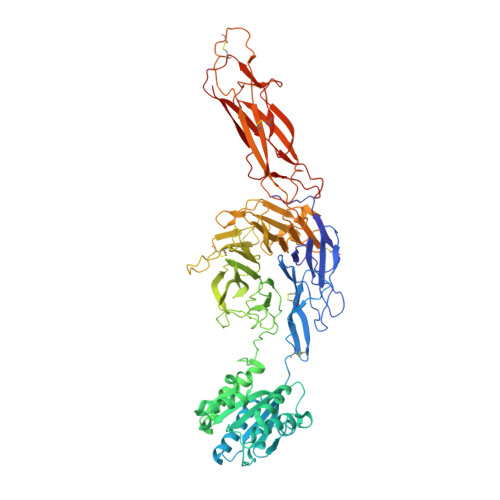
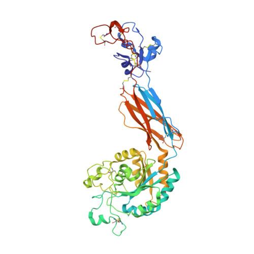
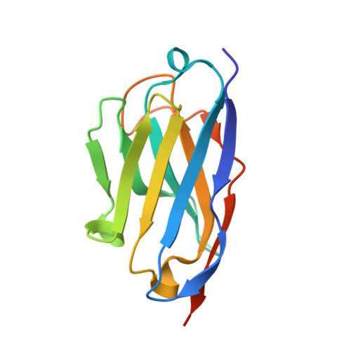
Structural insights into the function-modulating effects of nanobody binding to the integrin receptor alpha M beta 2.

Jensen, R.K.Pedersen, H.Lorentzen, J.Laursen, N.S.Vorup-Jensen, T.Andersen, G.R.

(2022) J Biol Chem 298: 102168-102168

  • DOI: https://doi.org/10.1016/j.jbc.2022.102168
  • Primary Citation of Related Structures:  
    7NP9, 7P2D

  • PubMed Abstract: 

    The integrin receptor α M β 2 mediates phagocytosis of complement-opsonized objects, adhesion to the extracellular matrix, and transendothelial migration of leukocytes. However, the mechanistic aspects of α M β 2 signaling upon ligand binding are unclear. Here, we present the first atomic structure of the human α M β 2 headpiece fragment in complex with the nanobody (Nb) hCD11bNb1 at a resolution of 3.2 Å. We show that the receptor headpiece adopts the closed conformation expected to exhibit low ligand affinity. The crystal structure indicates that in the R77H α M variant, associated with systemic lupus erythematosus, the modified allosteric relationship between ligand binding and integrin outside-inside signaling is due to subtle conformational effects transmitted over a distance of 40 Å. Furthermore, we found the Nb binds to the αI domain of the α M subunit in an Mg 2+ -independent manner with low nanomolar affinity. Biochemical and biophysical experiments with purified proteins demonstrated that the Nb acts as a competitive inhibitor through steric hindrance exerted on the thioester domain of complement component iC3b attempting to bind the α M subunit. Surprisingly, we show that the Nb stimulates the interaction of cell-bound α M β 2 with iC3b, suggesting that it may represent a novel high-affinity proteinaceous α M β 2 -specific agonist. Taken together, our data suggest that the iC3b-α M β 2 complex may be more dynamic than predicted from the crystal structure of the core complex. We propose a model based on the conformational spectrum of the receptor to reconcile these observations regarding the functional consequences of hCD11bNb1 binding to α M β 2 .


  • Organizational Affiliation

    Department of Molecular Biology and Genetics, Aarhus University, Denmark.


Macromolecules
Find similar proteins by:  (by identity cutoff)  |  3D Structure
Entity ID: 1
MoleculeChains Sequence LengthOrganismDetailsImage
Isoform 2 of Integrin alpha-M763Homo sapiensMutation(s): 0 
Gene Names: ITGAMCD11BCR3A
UniProt & NIH Common Fund Data Resources
Find proteins for P11215 (Homo sapiens)
Explore P11215 
Go to UniProtKB:  P11215
PHAROS:  P11215
GTEx:  ENSG00000169896 
Entity Groups  
Sequence Clusters30% Identity50% Identity70% Identity90% Identity95% Identity100% Identity
UniProt GroupP11215
Sequence Annotations
Expand
  • Reference Sequence
Find similar proteins by:  (by identity cutoff)  |  3D Structure
Entity ID: 2
MoleculeChains Sequence LengthOrganismDetailsImage
Integrin beta473Homo sapiensMutation(s): 0 
Gene Names: ITGB2
UniProt & NIH Common Fund Data Resources
Find proteins for P05107 (Homo sapiens)
Explore P05107 
Go to UniProtKB:  P05107
PHAROS:  P05107
GTEx:  ENSG00000160255 
Entity Groups  
Sequence Clusters30% Identity50% Identity70% Identity90% Identity95% Identity100% Identity
UniProt GroupP05107
Sequence Annotations
Expand
  • Reference Sequence
Find similar proteins by:  (by identity cutoff)  |  3D Structure
Entity ID: 3
MoleculeChains Sequence LengthOrganismDetailsImage
hCD11bNb1 nanobody129Lama glamaMutation(s): 0 
Entity Groups  
Sequence Clusters30% Identity50% Identity70% Identity90% Identity95% Identity100% Identity
Sequence Annotations
Expand
  • Reference Sequence
Oligosaccharides

Help

Entity ID: 4
MoleculeChains Length2D Diagram Glycosylation3D Interactions
2-acetamido-2-deoxy-beta-D-glucopyranose-(1-4)-2-acetamido-2-deoxy-beta-D-glucopyranose
D, E, F, G
2N-Glycosylation
Glycosylation Resources
GlyTouCan:  G42666HT
GlyCosmos:  G42666HT
GlyGen:  G42666HT
Experimental Data & Validation

Experimental Data

  • Method: X-RAY DIFFRACTION
  • Resolution: 3.20 Å
  • R-Value Free: 0.295 
  • R-Value Work: 0.261 
  • R-Value Observed: 0.262 
  • Space Group: P 31 2 1
Unit Cell:
Length ( Å )Angle ( ˚ )
a = 114.1α = 90
b = 114.1β = 90
c = 250.12γ = 120
Software Package:
Software NamePurpose
PHENIXrefinement
XDSdata reduction
XSCALEdata scaling
PHASERphasing

Structure Validation

View Full Validation Report


Ligand Structure Quality Assessment 


Entry History & Funding Information

Deposition Data


Funding OrganizationLocationGrant Number
LundbeckfondenDenmarkR155-2015-2666
Danish Council for Independent ResearchDenmark4181-00137
Novo Nordisk FoundationDenmarkNNF18OC0052105

Revision History  (Full details and data files)

  • Version 1.0: 2022-05-04
    Type: Initial release
  • Version 1.1: 2022-10-05
    Changes: Database references
  • Version 1.2: 2024-01-31
    Changes: Data collection, Refinement description