7XU4

Structure of SARS-CoV-2 D614G Spike Protein with Engineered x3 Disulfide (x3(D427C, V987C) and single Arg S1/S2 cleavage site), Locked-2 Conformation


Experimental Data Snapshot

  • Method: ELECTRON MICROSCOPY
  • Resolution: 3.20 Å
  • Aggregation State: PARTICLE 
  • Reconstruction Method: SINGLE PARTICLE 

wwPDB Validation   3D Report Full Report


This is version 1.1 of the entry. See complete history


Literature

Engineered disulfide reveals structural dynamics of locked SARS-CoV-2 spike.

Qu, K.Chen, Q.Ciazynska, K.A.Liu, B.Zhang, X.Wang, J.He, Y.Guan, J.He, J.Liu, T.Zhang, X.Carter, A.P.Xiong, X.Briggs, J.A.G.

(2022) PLoS Pathog 18: e1010583-e1010583

  • DOI: https://doi.org/10.1371/journal.ppat.1010583
  • Primary Citation of Related Structures:  
    7XTZ, 7XU0, 7XU1, 7XU2, 7XU3, 7XU4, 7XU5, 7XU6

  • PubMed Abstract: 

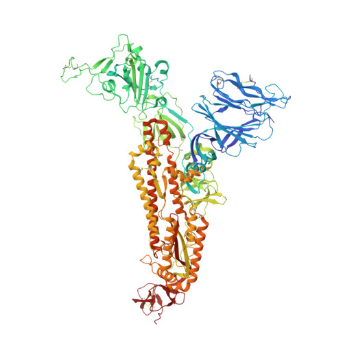
    The spike (S) protein of SARS-CoV-2 has been observed in three distinct pre-fusion conformations: locked, closed and open. Of these, the function of the locked conformation remains poorly understood. Here we engineered a SARS-CoV-2 S protein construct "S-R/x3" to arrest SARS-CoV-2 spikes in the locked conformation by a disulfide bond. Using this construct we determined high-resolution structures confirming that the x3 disulfide bond has the ability to stabilize the otherwise transient locked conformations. Structural analyses reveal that wild-type SARS-CoV-2 spike can adopt two distinct locked-1 and locked-2 conformations. For the D614G spike, based on which all variants of concern were evolved, only the locked-2 conformation was observed. Analysis of the structures suggests that rigidified domain D in the locked conformations interacts with the hinge to domain C and thereby restrains RBD movement. Structural change in domain D correlates with spike conformational change. We propose that the locked-1 and locked-2 conformations of S are present in the acidic high-lipid cellular compartments during virus assembly and egress. In this model, release of the virion into the neutral pH extracellular space would favour transition to the closed or open conformations. The dynamics of this transition can be altered by mutations that modulate domain D structure, as is the case for the D614G mutation, leading to changes in viral fitness. The S-R/x3 construct provides a tool for the further structural and functional characterization of the locked conformations of S, as well as how sequence changes might alter S assembly and regulation of receptor binding domain dynamics.


  • Organizational Affiliation

    Structural Studies Division, Medical Research Council Laboratory of Molecular Biology, Cambridge, United Kingdom.


Macromolecules
Find similar proteins by:  (by identity cutoff)  |  3D Structure
Entity ID: 1
MoleculeChains Sequence LengthOrganismDetailsImage
Spike glycoproteinA,
B [auth C],
C [auth B]
1,127Severe acute respiratory syndrome coronavirus 2Mutation(s): 3 
Gene Names: S2
UniProt
Find proteins for P0DTC2 (Severe acute respiratory syndrome coronavirus 2)
Explore P0DTC2 
Go to UniProtKB:  P0DTC2
Entity Groups  
Sequence Clusters30% Identity50% Identity70% Identity90% Identity95% Identity100% Identity
UniProt GroupP0DTC2
Sequence Annotations
Expand
  • Reference Sequence
Oligosaccharides

Help

Entity ID: 2
MoleculeChains Length2D Diagram Glycosylation3D Interactions
2-acetamido-2-deoxy-beta-D-glucopyranose-(1-4)-2-acetamido-2-deoxy-beta-D-glucopyranose
D, E, F, G, H
D, E, F, G, H, I, J, K, L, M, N, O
2N-Glycosylation
Glycosylation Resources
GlyTouCan:  G42666HT
GlyCosmos:  G42666HT
GlyGen:  G42666HT
Small Molecules
Ligands 3 Unique
IDChains Name / Formula / InChI Key2D Diagram3D Interactions
BLA
Query on BLA

Download Ideal Coordinates CCD File 
CA [auth A],
HB [auth B],
RA [auth C]
BILIVERDINE IX ALPHA
C33 H34 N4 O6
GWZYPXHJIZCRAJ-SRVCBVSDSA-N
EIC
Query on EIC

Download Ideal Coordinates CCD File 
DA [auth A],
SA [auth C],
TA [auth B]
LINOLEIC ACID
C18 H32 O2
OYHQOLUKZRVURQ-HZJYTTRNSA-N
NAG
Query on NAG

Download Ideal Coordinates CCD File 
AA [auth A]
AB [auth B]
BA [auth A]
BB [auth B]
CB [auth B]
AA [auth A],
AB [auth B],
BA [auth A],
BB [auth B],
CB [auth B],
DB [auth B],
EA [auth C],
EB [auth B],
FA [auth C],
FB [auth B],
GA [auth C],
GB [auth B],
HA [auth C],
IA [auth C],
JA [auth C],
KA [auth C],
LA [auth C],
MA [auth C],
NA [auth C],
OA [auth C],
P [auth A],
PA [auth C],
Q [auth A],
QA [auth C],
R [auth A],
S [auth A],
T [auth A],
U [auth A],
UA [auth B],
V [auth A],
VA [auth B],
W [auth A],
WA [auth B],
X [auth A],
XA [auth B],
Y [auth A],
YA [auth B],
Z [auth A],
ZA [auth B]
2-acetamido-2-deoxy-beta-D-glucopyranose
C8 H15 N O6
OVRNDRQMDRJTHS-FMDGEEDCSA-N
Experimental Data & Validation

Experimental Data

  • Method: ELECTRON MICROSCOPY
  • Resolution: 3.20 Å
  • Aggregation State: PARTICLE 
  • Reconstruction Method: SINGLE PARTICLE 
EM Software:
TaskSoftware PackageVersion
RECONSTRUCTIONRELION3.1

Structure Validation

View Full Validation Report


Entry History & Funding Information

Deposition Data


Funding OrganizationLocationGrant Number
European Research Council (ERC)European UnionERC-CoG-648432
Medical Research Council (MRC, United Kingdom)United KingdomMC_UP_A025_1011
Medical Research Council (MRC, United Kingdom)United KingdomMC_UP_1201/16

Revision History  (Full details and data files)

  • Version 1.0: 2022-07-20
    Type: Initial release
  • Version 1.1: 2022-08-17
    Changes: Database references