7UIA

Crystal structure of BoNT/E receptor binding domain in complex with SV2 and VHH


Experimental Data Snapshot

  • Method: X-RAY DIFFRACTION
  • Resolution: 2.59 Å
  • R-Value Free: 0.249 
  • R-Value Work: 0.226 
  • R-Value Observed: 0.227 

wwPDB Validation   3D Report Full Report


Ligand Structure Quality Assessment 


This is version 1.2 of the entry. See complete history


Literature

Structural basis for botulinum neurotoxin E recognition of synaptic vesicle protein 2.

Liu, Z.Lee, P.G.Krez, N.Lam, K.H.Liu, H.Przykopanski, A.Chen, P.Yao, G.Zhang, S.Tremblay, J.M.Perry, K.Shoemaker, C.B.Rummel, A.Dong, M.Jin, R.

(2023) Nat Commun 14: 2338-2338

  • DOI: https://doi.org/10.1038/s41467-023-37860-8
  • Primary Citation of Related Structures:  
    7UIA, 7UIB, 7UIE

  • PubMed Abstract: 

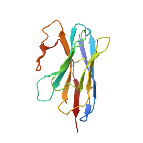
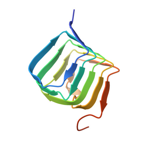
    Botulinum neurotoxin E (BoNT/E) is one of the major causes of human botulism and paradoxically also a promising therapeutic agent. Here we determined the co-crystal structures of the receptor-binding domain of BoNT/E (H C E) in complex with its neuronal receptor synaptic vesicle glycoprotein 2A (SV2A) and a nanobody that serves as a ganglioside surrogate. These structures reveal that the protein-protein interactions between H C E and SV2 provide the crucial location and specificity information for H C E to recognize SV2A and SV2B, but not the closely related SV2C. At the same time, H C E exploits a separated sialic acid-binding pocket to mediate recognition of an N-glycan of SV2. Structure-based mutagenesis and functional studies demonstrate that both the protein-protein and protein-glycan associations are essential for SV2A-mediated cell entry of BoNT/E and for its potent neurotoxicity. Our studies establish the structural basis to understand the receptor-specificity of BoNT/E and to engineer BoNT/E variants for new clinical applications.


  • Organizational Affiliation

    Department of Physiology and Biophysics, University of California, Irvine, Irvine, CA, 92697, USA.


Macromolecules
Find similar proteins by:  (by identity cutoff)  |  3D Structure
Entity ID: 1
MoleculeChains Sequence LengthOrganismDetailsImage
VHH-G6A [auth B],
C [auth E]
129Vicugna pacosMutation(s): 0 
Entity Groups  
Sequence Clusters30% Identity50% Identity70% Identity90% Identity95% Identity100% Identity
Sequence Annotations
Expand
  • Reference Sequence
Find similar proteins by:  (by identity cutoff)  |  3D Structure
Entity ID: 2
MoleculeChains Sequence LengthOrganismDetailsImage
SV2AcB [auth C],
D [auth F]
105Homo sapiensMutation(s): 0 
UniProt & NIH Common Fund Data Resources
Find proteins for Q496J9 (Homo sapiens)
Explore Q496J9 
Go to UniProtKB:  Q496J9
PHAROS:  Q496J9
GTEx:  ENSG00000122012 
Entity Groups  
Sequence Clusters30% Identity50% Identity70% Identity90% Identity95% Identity100% Identity
UniProt GroupQ496J9
Sequence Annotations
Expand
  • Reference Sequence
Find similar proteins by:  (by identity cutoff)  |  3D Structure
Entity ID: 3
MoleculeChains Sequence LengthOrganismDetailsImage
Neurotoxin type EE [auth D],
F [auth A]
407Clostridium botulinumMutation(s): 0 
UniProt
Find proteins for Q00496 (Clostridium botulinum)
Explore Q00496 
Go to UniProtKB:  Q00496
Entity Groups  
Sequence Clusters30% Identity50% Identity70% Identity90% Identity95% Identity100% Identity
UniProt GroupQ00496
Sequence Annotations
Expand
  • Reference Sequence
Oligosaccharides

Help

Entity ID: 4
MoleculeChains Length2D Diagram Glycosylation3D Interactions
2-acetamido-2-deoxy-beta-D-glucopyranose-(1-4)-2-acetamido-2-deoxy-beta-D-glucopyranose
G, H
2N-Glycosylation
Glycosylation Resources
GlyTouCan:  G42666HT
GlyCosmos:  G42666HT
GlyGen:  G42666HT
Small Molecules
Ligands 3 Unique
IDChains Name / Formula / InChI Key2D Diagram3D Interactions
NAG (Subject of Investigation/LOI)
Query on NAG

Download Ideal Coordinates CCD File 
I [auth C]
J [auth C]
M [auth F]
N [auth F]
O [auth F]
I [auth C],
J [auth C],
M [auth F],
N [auth F],
O [auth F],
P [auth F]
2-acetamido-2-deoxy-beta-D-glucopyranose
C8 H15 N O6
OVRNDRQMDRJTHS-FMDGEEDCSA-N
PEG
Query on PEG

Download Ideal Coordinates CCD File 
T [auth A]DI(HYDROXYETHYL)ETHER
C4 H10 O3
MTHSVFCYNBDYFN-UHFFFAOYSA-N
SO4
Query on SO4

Download Ideal Coordinates CCD File 
K [auth C]
L [auth C]
Q [auth F]
R [auth D]
S [auth D]
K [auth C],
L [auth C],
Q [auth F],
R [auth D],
S [auth D],
U [auth A],
V [auth A]
SULFATE ION
O4 S
QAOWNCQODCNURD-UHFFFAOYSA-L
Experimental Data & Validation

Experimental Data

  • Method: X-RAY DIFFRACTION
  • Resolution: 2.59 Å
  • R-Value Free: 0.249 
  • R-Value Work: 0.226 
  • R-Value Observed: 0.227 
  • Space Group: C 2 2 21
Unit Cell:
Length ( Å )Angle ( ˚ )
a = 140.742α = 90
b = 172.335β = 90
c = 137.162γ = 90
Software Package:
Software NamePurpose
REFMACrefinement
XDSdata reduction
XDSdata scaling
PHENIXphasing

Structure Validation

View Full Validation Report


Ligand Structure Quality Assessment 


Entry History & Funding Information

Deposition Data


Funding OrganizationLocationGrant Number
National Institutes of Health/National Institute Of Allergy and Infectious Diseases (NIH/NIAID)United States--

Revision History  (Full details and data files)

  • Version 1.0: 2023-04-05
    Type: Initial release
  • Version 1.1: 2023-05-03
    Changes: Database references, Source and taxonomy, Structure summary
  • Version 1.2: 2023-10-25
    Changes: Data collection, Refinement description