7R3B

S-adenosylmethionine synthetase from Lactobacillus plantarum complexed with AMPPNP, methionine and SAM


Experimental Data Snapshot

  • Method: X-RAY DIFFRACTION
  • Resolution: 2.82 Å
  • R-Value Free: 0.275 
  • R-Value Work: 0.156 
  • R-Value Observed: 0.161 

wwPDB Validation   3D Report Full Report


Ligand Structure Quality Assessment 


This is version 1.1 of the entry. See complete history


Literature

Evolution of homo-oligomerization of methionine S-adenosyltransferases is replete with structure-function constrains.

Kleiner, D.Shapiro Tuchman, Z.Shmulevich, F.Shahar, A.Zarivach, R.Kosloff, M.Bershtein, S.

(2022) Protein Sci 31: e4352-e4352

  • DOI: https://doi.org/10.1002/pro.4352
  • Primary Citation of Related Structures:  
    7R2W, 7R3B

  • PubMed Abstract: 

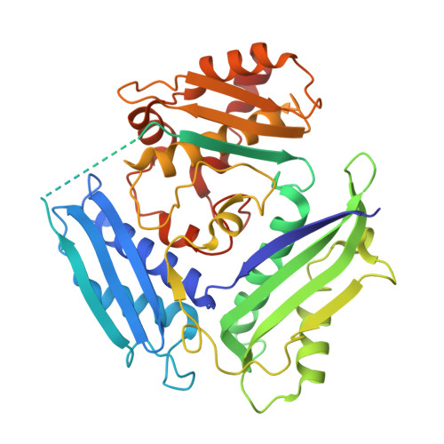
    Homomers are prevalent in bacterial proteomes, particularly among core metabolic enzymes. Homomerization is often key to function and regulation, and interfaces that facilitate the formation of homomeric enzymes are subject to intense evolutionary change. However, our understanding of the molecular mechanisms that drive evolutionary variation in homomeric complexes is still lacking. How is the diversification of protein interfaces linked to variation in functional regulation and structural integrity of homomeric complexes? To address this question, we studied quaternary structure evolution of bacterial methionine S-adenosyltransferases (MATs)-dihedral homotetramers formed along a large and conserved dimeric interface harboring two active sites, and a small, recently evolved, interdimeric interface. Here, we show that diversity in the physicochemical properties of small interfaces is directly linked to variability in the kinetic stability of MAT quaternary complexes and in modes of their functional regulation. Specifically, hydrophobic interactions within the small interface of Escherichia coli MAT render the functional homotetramer kinetically stable yet impose severe aggregation constraints on complex assembly. These constraints are alleviated by electrostatic interactions that accelerate dimer-dimer assembly. In contrast, Neisseria gonorrhoeae MAT adopts a nonfunctional dimeric state due to the low hydrophobicity of its small interface and the high flexibility of its active site loops, which perturbs small interface integrity. Remarkably, in the presence of methionine and ATP, N. gonorrhoeae MAT undergoes substrate-induced assembly into a functional tetrameric state. We suggest that evolution acts on the interdimeric interfaces of MATs to tailor the regulation of their activity and stability to unique organismal needs.


  • Organizational Affiliation

    Department of Life Sciences, Ben-Gurion University of the Negev, Beer-Sheva, Israel.


Macromolecules
Find similar proteins by:  (by identity cutoff)  |  3D Structure
Entity ID: 1
MoleculeChains Sequence LengthOrganismDetailsImage
S-adenosylmethionine synthase
A, B, C, D, E
A, B, C, D, E, F, G, H
401Lactiplantibacillus plantarumMutation(s): 0 
Gene Names: 
EC: 2.5.1.6
UniProt
Find proteins for Q88XB8 (Lactiplantibacillus plantarum (strain ATCC BAA-793 / NCIMB 8826 / WCFS1))
Explore Q88XB8 
Go to UniProtKB:  Q88XB8
Entity Groups  
Sequence Clusters30% Identity50% Identity70% Identity90% Identity95% Identity100% Identity
UniProt GroupQ88XB8
Sequence Annotations
Expand
  • Reference Sequence
Small Molecules
Ligands 6 Unique
IDChains Name / Formula / InChI Key2D Diagram3D Interactions
ANP (Subject of Investigation/LOI)
Query on ANP

Download Ideal Coordinates CCD File 
AA [auth F],
Q [auth C],
U [auth D],
Y [auth E]
PHOSPHOAMINOPHOSPHONIC ACID-ADENYLATE ESTER
C10 H17 N6 O12 P3
PVKSNHVPLWYQGJ-KQYNXXCUSA-N
SAM (Subject of Investigation/LOI)
Query on SAM

Download Ideal Coordinates CCD File 
GA [auth G],
JA [auth H]
S-ADENOSYLMETHIONINE
C15 H22 N6 O5 S
MEFKEPWMEQBLKI-FCKMPRQPSA-N
PPK (Subject of Investigation/LOI)
Query on PPK

Download Ideal Coordinates CCD File 
FA [auth G],
J [auth A],
LA [auth H],
O [auth B]
(DIPHOSPHONO)AMINOPHOSPHONIC ACID
H6 N O9 P3
PELPUMGXMYVGSQ-UHFFFAOYSA-N
PHE (Subject of Investigation/LOI)
Query on PHE

Download Ideal Coordinates CCD File 
I [auth A],
KA [auth H]
PHENYLALANINE
C9 H11 N O2
COLNVLDHVKWLRT-QMMMGPOBSA-N
K (Subject of Investigation/LOI)
Query on K

Download Ideal Coordinates CCD File 
EA [auth F],
N [auth A],
OA [auth H],
T [auth C],
X [auth D]
POTASSIUM ION
K
NPYPAHLBTDXSSS-UHFFFAOYSA-N
MG (Subject of Investigation/LOI)
Query on MG

Download Ideal Coordinates CCD File 
BA [auth F]
CA [auth F]
DA [auth F]
HA [auth G]
IA [auth G]
BA [auth F],
CA [auth F],
DA [auth F],
HA [auth G],
IA [auth G],
K [auth A],
L [auth A],
M [auth A],
MA [auth H],
NA [auth H],
P [auth B],
R [auth C],
S [auth C],
V [auth D],
W [auth D],
Z [auth E]
MAGNESIUM ION
Mg
JLVVSXFLKOJNIY-UHFFFAOYSA-N
Experimental Data & Validation

Experimental Data

  • Method: X-RAY DIFFRACTION
  • Resolution: 2.82 Å
  • R-Value Free: 0.275 
  • R-Value Work: 0.156 
  • R-Value Observed: 0.161 
  • Space Group: P 1
Unit Cell:
Length ( Å )Angle ( ˚ )
a = 58.426α = 93.82
b = 110.927β = 104.03
c = 112.661γ = 99.59
Software Package:
Software NamePurpose
REFMACrefinement
xia2data reduction
DIALSdata scaling
PHASERphasing

Structure Validation

View Full Validation Report


Ligand Structure Quality Assessment 


Entry History & Funding Information

Deposition Data


Funding OrganizationLocationGrant Number
United States - Israel Binational Science Foundation (BSF)United States2020640

Revision History  (Full details and data files)

  • Version 1.0: 2022-07-13
    Type: Initial release
  • Version 1.1: 2024-01-31
    Changes: Data collection, Refinement description