7QOT

Factor XI and Plasma Kallikrein apple domain structures reveals different kininogen bound complexes


Experimental Data Snapshot

  • Method: X-RAY DIFFRACTION
  • Resolution: 3.24 Å
  • R-Value Free: 0.284 
  • R-Value Work: 0.251 
  • R-Value Observed: 0.254 

wwPDB Validation   3D Report Full Report


Ligand Structure Quality Assessment 


This is version 1.1 of the entry. See complete history


Literature

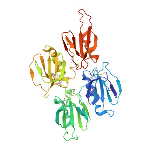
Plasma kallikrein structure reveals apple domain disc rotated conformation compared to factor XI.

Li, C.Voos, K.M.Pathak, M.Hall, G.McCrae, K.R.Dreveny, I.Li, R.Emsley, J.

(2019) J Thromb Haemost 17: 759-770

  • DOI: https://doi.org/10.1111/jth.14418
  • Primary Citation of Related Structures:  
    6I44, 6I58, 7QOT, 7QOX

  • PubMed Abstract: 

    Essentials Zymogen PK is activated to PKa and cleaves substrates kininogen and FXII contributing to bradykinin generation. Monomeric PKa and dimeric homologue FXI utilize the N-terminal apple domains to recruit substrates. A high-resolution 1.3 Å structure of full-length PKa reveals an active conformation of the protease and apple domains. The PKa protease and four-apple domain disc organization is 180° rotated compared to FXI. SUMMARY: Background Plasma prekallikrein (PK) and factor XI (FXI) are apple domain-containing serine proteases that when activated to PKa and FXIa cleave substrates kininogen, factor XII, and factor IX, respectively, directing plasma coagulation, bradykinin release, inflammation, and thrombosis pathways. Objective To investigate the three-dimensional structure of full-length PKa and perform a comparison with FXI. Methods A series of recombinant full-length PKa and FXI constructs and variants were developed and the crystal structures determined. Results and conclusions A 1.3 Å structure of full-length PKa reveals the protease domain positioned above a disc-shaped assemblage of four apple domains in an active conformation. A comparison with the homologous FXI structure reveals the intramolecular disulfide and structural differences in the apple 4 domain that prevents dimer formation in PK as opposed to FXI. Two latchlike loops (LL1 and LL2) extend from the PKa protease domain to form interactions with the apple 1 and apple 3 domains, respectively. A major unexpected difference in the PKa structure compared to FXI is the 180° disc rotation of the apple domains relative to the protease domain. This results in a switched configuration of the latch loops such that LL2 interacts and buries portions of the apple 3 domain in the FXI zymogen whereas in PKa LL2 interacts with the apple 1 domain. Hydrogen-deuterium exchange mass spectrometry on plasma purified human PK and PKa determined that regions of the apple 3 domain have increased surface exposure in PKa compared to the zymogen PK, suggesting conformational change upon activation.


  • Organizational Affiliation

    Centre for Biomolecular Sciences, School of Pharmacy, University of Nottingham, Nottingham, UK.


Macromolecules
Find similar proteins by:  (by identity cutoff)  |  3D Structure
Entity ID: 1
MoleculeChains Sequence LengthOrganismDetailsImage
Coagulation factor XIa heavy chain
A, B
358Homo sapiensMutation(s): 0 
Gene Names: F11
UniProt & NIH Common Fund Data Resources
Find proteins for P03951 (Homo sapiens)
Explore P03951 
Go to UniProtKB:  P03951
PHAROS:  P03951
GTEx:  ENSG00000088926 
Entity Groups  
Sequence Clusters30% Identity50% Identity70% Identity90% Identity95% Identity100% Identity
UniProt GroupP03951
Sequence Annotations
Expand
  • Reference Sequence
Find similar proteins by:  (by identity cutoff)  |  3D Structure
Entity ID: 2
MoleculeChains Sequence LengthOrganismDetailsImage
Kininogen-1 light chain
C, D
31Homo sapiensMutation(s): 0 
Gene Names: KNG1BDKKNG
UniProt & NIH Common Fund Data Resources
Find proteins for P01042 (Homo sapiens)
Explore P01042 
Go to UniProtKB:  P01042
PHAROS:  P01042
GTEx:  ENSG00000113889 
Entity Groups  
Sequence Clusters30% Identity50% Identity70% Identity90% Identity95% Identity100% Identity
UniProt GroupP01042
Sequence Annotations
Expand
  • Reference Sequence
Small Molecules
Ligands 2 Unique
IDChains Name / Formula / InChI Key2D Diagram3D Interactions
NAG (Subject of Investigation/LOI)
Query on NAG

Download Ideal Coordinates CCD File 
E [auth A],
F [auth A],
J [auth B]
2-acetamido-2-deoxy-beta-D-glucopyranose
C8 H15 N O6
OVRNDRQMDRJTHS-FMDGEEDCSA-N
SO4 (Subject of Investigation/LOI)
Query on SO4

Download Ideal Coordinates CCD File 
G [auth A]
H [auth A]
I [auth A]
K [auth B]
L [auth B]
G [auth A],
H [auth A],
I [auth A],
K [auth B],
L [auth B],
M [auth B]
SULFATE ION
O4 S
QAOWNCQODCNURD-UHFFFAOYSA-L
Experimental Data & Validation

Experimental Data

  • Method: X-RAY DIFFRACTION
  • Resolution: 3.24 Å
  • R-Value Free: 0.284 
  • R-Value Work: 0.251 
  • R-Value Observed: 0.254 
  • Space Group: P 61 2 2
Unit Cell:
Length ( Å )Angle ( ˚ )
a = 87.319α = 90
b = 87.319β = 90
c = 629.548γ = 120
Software Package:
Software NamePurpose
PHENIXrefinement
XDSdata reduction
Aimlessdata scaling
PHASERphasing
PDB_EXTRACTdata extraction

Structure Validation

View Full Validation Report


Ligand Structure Quality Assessment 


Entry History & Funding Information

Deposition Data


Funding OrganizationLocationGrant Number
British Heart FoundationUnited KingdomRG/12/9/29775

Revision History  (Full details and data files)

  • Version 1.0: 2023-01-18
    Type: Initial release
  • Version 1.1: 2024-01-31
    Changes: Data collection, Refinement description