7QLJ

Trans structure of rsKiiro Illuminated at 290 K


Experimental Data Snapshot

  • Method: X-RAY DIFFRACTION
  • Resolution: 1.02 Å
  • R-Value Free: 0.137 
  • R-Value Work: 0.119 

wwPDB Validation   3D Report Full Report


This is version 4.0 of the entry. See complete history


Literature

Optical control of ultrafast structural dynamics in a fluorescent protein.

Hutchison, C.D.M.Baxter, J.M.Fitzpatrick, A.Dorlhiac, G.Fadini, A.Perrett, S.Maghlaoui, K.Lefevre, S.B.Cordon-Preciado, V.Ferreira, J.L.Chukhutsina, V.U.Garratt, D.Barnard, J.Galinis, G.Glencross, F.Morgan, R.M.Stockton, S.Taylor, B.Yuan, L.Romei, M.G.Lin, C.Y.Marangos, J.P.Schmidt, M.Chatrchyan, V.Buckup, T.Morozov, D.Park, J.Park, S.Eom, I.Kim, M.Jang, D.Choi, H.Hyun, H.Park, G.Nango, E.Tanaka, R.Owada, S.Tono, K.DePonte, D.P.Carbajo, S.Seaberg, M.Aquila, A.Boutet, S.Barty, A.Iwata, S.Boxer, S.G.Groenhof, G.van Thor, J.J.

(2023) Nat Chem 15: 1607-1615

  • DOI: https://doi.org/10.1038/s41557-023-01275-1
  • Primary Citation of Related Structures:  
    7QLI, 7QLJ, 7QLK, 7QLL, 7QLM, 7QLN, 7QLO

  • PubMed Abstract: 

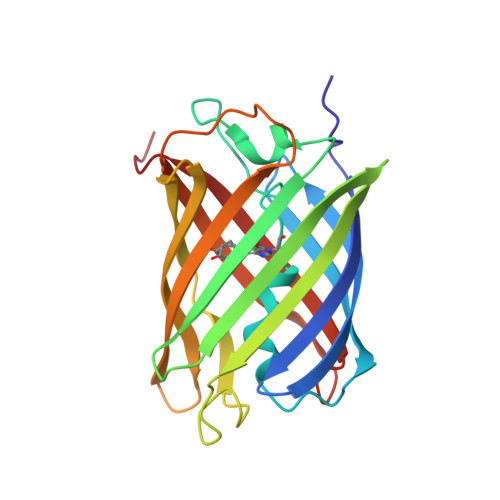
    The photoisomerization reaction of a fluorescent protein chromophore occurs on the ultrafast timescale. The structural dynamics that result from femtosecond optical excitation have contributions from vibrational and electronic processes and from reaction dynamics that involve the crossing through a conical intersection. The creation and progression of the ultrafast structural dynamics strongly depends on optical and molecular parameters. When using X-ray crystallography as a probe of ultrafast dynamics, the origin of the observed nuclear motions is not known. Now, high-resolution pump-probe X-ray crystallography reveals complex sub-ångström, ultrafast motions and hydrogen-bonding rearrangements in the active site of a fluorescent protein. However, we demonstrate that the measured motions are not part of the photoisomerization reaction but instead arise from impulsively driven coherent vibrational processes in the electronic ground state. A coherent-control experiment using a two-colour and two-pulse optical excitation strongly amplifies the X-ray crystallographic difference density, while it fully depletes the photoisomerization process. A coherent control mechanism was tested and confirmed the wave packets assignment.


  • Organizational Affiliation

    Department of Life Sciences, Faculty of Natural Sciences, Imperial College London, London, UK.


Macromolecules
Find similar proteins by:  (by identity cutoff)  |  3D Structure
Entity ID: 1
MoleculeChains Sequence LengthOrganismDetailsImage
rsKiiro220Lobophyllia hemprichiiMutation(s): 12 
UniProt
Find proteins for Q5S6Z9 (Lobophyllia hemprichii)
Explore Q5S6Z9 
Go to UniProtKB:  Q5S6Z9
Entity Groups  
Sequence Clusters30% Identity50% Identity70% Identity90% Identity95% Identity100% Identity
UniProt GroupQ5S6Z9
Sequence Annotations
Expand
  • Reference Sequence
Small Molecules
Ligands 1 Unique
IDChains Name / Formula / InChI Key2D Diagram3D Interactions
SO4
Query on SO4

Download Ideal Coordinates CCD File 
B [auth A]SULFATE ION
O4 S
QAOWNCQODCNURD-UHFFFAOYSA-L
Modified Residues  1 Unique
IDChains TypeFormula2D DiagramParent
PIA
Query on PIA
A
L-PEPTIDE LINKINGC14 H15 N3 O4ALA, TYR, GLY
Experimental Data & Validation

Experimental Data

  • Method: X-RAY DIFFRACTION
  • Resolution: 1.02 Å
  • R-Value Free: 0.137 
  • R-Value Work: 0.119 
  • Space Group: P 21 21 21
Unit Cell:
Length ( Å )Angle ( ˚ )
a = 38.747α = 90
b = 73.524β = 90
c = 78.106γ = 90
Software Package:
Software NamePurpose
REFMACrefinement
autoPROCdata reduction
STARANISOdata scaling
REFMACphasing

Structure Validation

View Full Validation Report


Entry History & Funding Information

Deposition Data


Funding OrganizationLocationGrant Number
Biotechnology and Biological Sciences Research Council (BBSRC)United KingdomBB/P00752X/1

Revision History  (Full details and data files)

  • Version 1.0: 2022-11-09
    Type: Initial release
  • Version 1.1: 2022-11-16
    Changes: Database references
  • Version 1.2: 2022-11-30
    Changes: Database references
  • Version 2.0: 2022-12-28
    Changes: Advisory, Atomic model, Author supporting evidence, Data collection, Database references, Derived calculations, Non-polymer description, Polymer sequence, Refinement description, Structure summary
  • Version 3.0: 2023-04-05
    Changes: Atomic model, Database references, Source and taxonomy
  • Version 3.1: 2023-08-16
    Changes: Data collection, Database references
  • Version 3.2: 2023-08-23
    Changes: Database references, Structure summary
  • Version 4.0: 2023-11-15
    Changes: Atomic model, Data collection, Database references