7Q6I

Vibrio maritimus FtsA 1-396 ATP and FtsN 1-29, bent tetramers in double filament arrangement


Experimental Data Snapshot

  • Method: X-RAY DIFFRACTION
  • Resolution: 3.60 Å
  • R-Value Free: 0.287 
  • R-Value Work: 0.241 
  • R-Value Observed: 0.243 

wwPDB Validation   3D Report Full Report


Ligand Structure Quality Assessment 


This is version 1.4 of the entry. See complete history


Literature

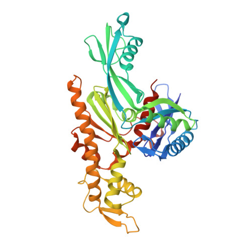
Bacterial divisome protein FtsA forms curved antiparallel double filaments when binding to FtsN.

Nierhaus, T.McLaughlin, S.H.Burmann, F.Kureisaite-Ciziene, D.Maslen, S.L.Skehel, J.M.Yu, C.W.H.Freund, S.M.V.Funke, L.F.H.Chin, J.W.Lowe, J.

(2022) Nat Microbiol 7: 1686-1701

  • DOI: https://doi.org/10.1038/s41564-022-01206-9
  • Primary Citation of Related Structures:  
    7Q6D, 7Q6F, 7Q6G, 7Q6I

  • PubMed Abstract: 

    During bacterial cell division, filaments of tubulin-like FtsZ form the Z-ring, which is the cytoplasmic scaffold for divisome assembly. In Escherichia coli, the actin homologue FtsA anchors the Z-ring to the membrane and recruits divisome components, including bitopic FtsN. FtsN regulates the periplasmic peptidoglycan synthase FtsWI. To characterize how FtsA regulates FtsN, we applied electron microscopy to show that E. coli FtsA forms antiparallel double filaments on lipid monolayers when bound to the cytoplasmic tail of FtsN. Using X-ray crystallography, we demonstrate that Vibrio maritimus FtsA crystallizes as an equivalent double filament. We identified an FtsA-FtsN interaction site in the IA-IC interdomain cleft of FtsA using X-ray crystallography and confirmed that FtsA forms double filaments in vivo by site-specific cysteine cross-linking. FtsA-FtsN double filaments reconstituted in or on liposomes prefer negative Gaussian curvature, like those of MreB, the actin-like protein of the elongasome. We propose that curved antiparallel FtsA double filaments together with treadmilling FtsZ filaments organize septal peptidoglycan synthesis in the division plane.


  • Organizational Affiliation

    MRC Laboratory of Molecular Biology, Cambridge, UK.


Macromolecules
Find similar proteins by:  (by identity cutoff)  |  3D Structure
Entity ID: 1
MoleculeChains Sequence LengthOrganismDetailsImage
Cell division protein FtsA
A, B, C, D, E
A, B, C, D, E, F, G, H, I, J, K, L, M, N, O, P
396Vibrio maritimusMutation(s): 0 
Gene Names: ftsAJCM19240_1885
UniProt
Find proteins for A0A090T942 (Vibrio maritimus)
Explore A0A090T942 
Go to UniProtKB:  A0A090T942
Entity Groups  
Sequence Clusters30% Identity50% Identity70% Identity90% Identity95% Identity100% Identity
UniProt GroupA0A090T942
Sequence Annotations
Expand
  • Reference Sequence
Find similar proteins by:  (by identity cutoff)  |  3D Structure
Entity ID: 2
MoleculeChains Sequence LengthOrganismDetailsImage
Cell division protein FtsN (polyAla model)Q [auth X],
R [auth Y]
37Vibrio maritimusMutation(s): 0 
Entity Groups  
Sequence Clusters30% Identity50% Identity70% Identity90% Identity95% Identity100% Identity
Sequence Annotations
Expand
  • Reference Sequence
Small Molecules
Ligands 2 Unique
IDChains Name / Formula / InChI Key2D Diagram3D Interactions
ATP (Subject of Investigation/LOI)
Query on ATP

Download Ideal Coordinates CCD File 
AA [auth E]
CA [auth F]
EA [auth G]
GA [auth H]
IA [auth I]
AA [auth E],
CA [auth F],
EA [auth G],
GA [auth H],
IA [auth I],
KA [auth J],
MA [auth K],
OA [auth L],
QA [auth M],
S [auth A],
SA [auth N],
U [auth B],
UA [auth O],
W [auth C],
WA [auth P],
Y [auth D]
ADENOSINE-5'-TRIPHOSPHATE
C10 H16 N5 O13 P3
ZKHQWZAMYRWXGA-KQYNXXCUSA-N
MG
Query on MG

Download Ideal Coordinates CCD File 
BA [auth E]
DA [auth F]
FA [auth G]
HA [auth H]
JA [auth I]
BA [auth E],
DA [auth F],
FA [auth G],
HA [auth H],
JA [auth I],
LA [auth J],
NA [auth K],
PA [auth L],
RA [auth M],
T [auth A],
TA [auth N],
V [auth B],
VA [auth O],
X [auth C],
XA [auth P],
Z [auth D]
MAGNESIUM ION
Mg
JLVVSXFLKOJNIY-UHFFFAOYSA-N
Experimental Data & Validation

Experimental Data

  • Method: X-RAY DIFFRACTION
  • Resolution: 3.60 Å
  • R-Value Free: 0.287 
  • R-Value Work: 0.241 
  • R-Value Observed: 0.243 
  • Space Group: P 1 21 1
Unit Cell:
Length ( Å )Angle ( ˚ )
a = 98.692α = 90
b = 119.993β = 93.76
c = 329.788γ = 90
Software Package:
Software NamePurpose
PHENIXrefinement
REFMACrefinement
Aimlessdata scaling
PDB_EXTRACTdata extraction
XDSdata reduction
PHASERphasing

Structure Validation

View Full Validation Report


Ligand Structure Quality Assessment 


Entry History & Funding Information

Deposition Data


Funding OrganizationLocationGrant Number
Wellcome TrustUnited Kingdom202754/Z/16/Z
Medical Research Council (MRC, United Kingdom)United KingdomU105184326

Revision History  (Full details and data files)

  • Version 1.0: 2022-09-21
    Type: Initial release
  • Version 1.1: 2022-09-28
    Changes: Database references
  • Version 1.2: 2022-10-05
    Changes: Database references
  • Version 1.3: 2022-10-12
    Changes: Database references
  • Version 1.4: 2024-01-31
    Changes: Data collection, Refinement description