7MN8

Structure of the HER2/HER3/NRG1b Heterodimer Extracellular Domain bound to Trastuzumab Fab


Experimental Data Snapshot

  • Method: ELECTRON MICROSCOPY
  • Resolution: 3.45 Å
  • Aggregation State: PARTICLE 
  • Reconstruction Method: SINGLE PARTICLE 

wwPDB Validation   3D Report Full Report


This is version 1.2 of the entry. See complete history


Literature

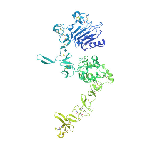
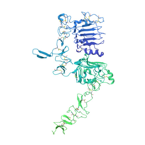
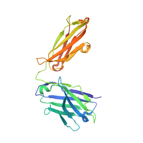
Structures of the HER2-HER3-NRG1 beta complex reveal a dynamic dimer interface.

Diwanji, D.Trenker, R.Thaker, T.M.Wang, F.Agard, D.A.Verba, K.A.Jura, N.

(2021) Nature 600: 339-343

  • DOI: https://doi.org/10.1038/s41586-021-04084-z
  • Primary Citation of Related Structures:  
    7MN5, 7MN6, 7MN8

  • PubMed Abstract: 

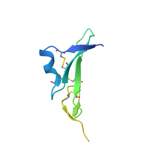
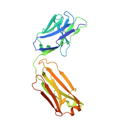
    Human epidermal growth factor receptor 2 (HER2) and HER3 form a potent pro-oncogenic heterocomplex 1-3 upon binding of growth factor neuregulin-1β (NRG1β). The mechanism by which HER2 and HER3 interact remains unknown in the absence of any structures of the complex. Here we isolated the NRG1β-bound near full-length HER2-HER3 dimer and, using cryo-electron microscopy, reconstructed the extracellulardomain module, revealing unexpected dynamics at the HER2-HER3 dimerization interface. We show that the dimerization arm of NRG1β-bound HER3 is unresolved because the apo HER2 monomer does not undergo a ligand-induced conformational change needed to establish a HER3 dimerization arm-binding pocket. In a structure of the oncogenic extracellular domain mutant HER2(S310F), we observe a compensatory interaction with the HER3 dimerization arm that stabilizes the dimerization interface. Both HER2-HER3 and HER2(S310F)-HER3 retain the capacity to bind to the HER2-directed therapeutic antibody trastuzumab, but the mutant complex does not bind to pertuzumab. Our structure of the HER2(S310F)-HER3-NRG1β-trastuzumab Fab complex reveals that the receptor dimer undergoes a conformational change to accommodate trastuzumab. Thus, similar to oncogenic mutations, therapeutic agents exploit the intrinsic dynamics of the HER2-HER3 heterodimer. The unique features of a singly liganded HER2-HER3 heterodimer underscore the allosteric sensing of ligand occupancy by the dimerization interface and explain why extracellular domains of HER2 do not homo-associate via a canonical active dimer interface.


  • Organizational Affiliation

    Cardiovascular Research Institute, University of California San Francisco, San Francisco, CA, USA.


Macromolecules
Find similar proteins by:  (by identity cutoff)  |  3D Structure
Entity ID: 1
MoleculeChains Sequence LengthOrganismDetailsImage
Receptor tyrosine-protein kinase erbB-31,066Homo sapiensMutation(s): 0 
Gene Names: ERBB3HER3
EC: 2.7.10.1
UniProt & NIH Common Fund Data Resources
Find proteins for P21860 (Homo sapiens)
Explore P21860 
Go to UniProtKB:  P21860
PHAROS:  P21860
GTEx:  ENSG00000065361 
Entity Groups  
Sequence Clusters30% Identity50% Identity70% Identity90% Identity95% Identity100% Identity
UniProt GroupP21860
Sequence Annotations
Expand
  • Reference Sequence
Find similar proteins by:  (by identity cutoff)  |  3D Structure
Entity ID: 2
MoleculeChains Sequence LengthOrganismDetailsImage
Isoform 6 of Pro-neuregulin-1, membrane-bound isoformB [auth H]87Homo sapiensMutation(s): 0 
Gene Names: NRG1GGFHGLHRGANDFSMDF
UniProt & NIH Common Fund Data Resources
Find proteins for Q02297 (Homo sapiens)
Explore Q02297 
Go to UniProtKB:  Q02297
PHAROS:  Q02297
GTEx:  ENSG00000157168 
Entity Groups  
Sequence Clusters30% Identity50% Identity70% Identity90% Identity95% Identity100% Identity
UniProt GroupQ02297
Sequence Annotations
Expand
  • Reference Sequence
Find similar proteins by:  (by identity cutoff)  |  3D Structure
Entity ID: 3
MoleculeChains Sequence LengthOrganismDetailsImage
Receptor tyrosine-protein kinase erbB-2,Maltose/maltodextrin-binding periplasmic proteinC [auth B]1,455Homo sapiensEscherichia coli K-12
This entity is chimeric
Mutation(s): 1 
Gene Names: ERBB2HER2MLN19NEUNGLmalEb4034JW3994
EC: 2.7.10.1
UniProt & NIH Common Fund Data Resources
Find proteins for P0AEX9 (Escherichia coli (strain K12))
Explore P0AEX9 
Go to UniProtKB:  P0AEX9
Find proteins for P04626 (Homo sapiens)
Explore P04626 
Go to UniProtKB:  P04626
PHAROS:  P04626
GTEx:  ENSG00000141736 
Entity Groups  
Sequence Clusters30% Identity50% Identity70% Identity90% Identity95% Identity100% Identity
UniProt GroupsP04626P0AEX9
Sequence Annotations
Expand
  • Reference Sequence
Find similar proteins by:  (by identity cutoff)  |  3D Structure
Entity ID: 4
MoleculeChains Sequence LengthOrganismDetailsImage
Trastuzumab Fab Light ChainD [auth C]225Homo sapiensMutation(s): 0 
Entity Groups  
Sequence Clusters30% Identity50% Identity70% Identity90% Identity95% Identity100% Identity
Sequence Annotations
Expand
  • Reference Sequence
Find similar proteins by:  (by identity cutoff)  |  3D Structure
Entity ID: 5
MoleculeChains Sequence LengthOrganismDetailsImage
Trastuzumab Fab Heavy ChainE [auth D]237Homo sapiensMutation(s): 0 
Entity Groups  
Sequence Clusters30% Identity50% Identity70% Identity90% Identity95% Identity100% Identity
Sequence Annotations
Expand
  • Reference Sequence
Oligosaccharides

Help

Entity ID: 6
MoleculeChains Length2D Diagram Glycosylation3D Interactions
2-acetamido-2-deoxy-beta-D-glucopyranose-(1-4)-2-acetamido-2-deoxy-beta-D-glucopyranoseF [auth E],
G [auth F]
2N/AN-Glycosylation
Glycosylation Resources
GlyTouCan:  G42666HT
GlyCosmos:  G42666HT
GlyGen:  G42666HT
Entity ID: 7
MoleculeChains Length2D Diagram Glycosylation3D Interactions
alpha-D-mannopyranose-(1-3)-beta-D-mannopyranose-(1-4)-2-acetamido-2-deoxy-beta-D-glucopyranose-(1-4)-2-acetamido-2-deoxy-beta-D-glucopyranoseH [auth G]4N/AN-Glycosylation
Glycosylation Resources
GlyTouCan:  G81315DD
GlyCosmos:  G81315DD
GlyGen:  G81315DD
Experimental Data & Validation

Experimental Data

  • Method: ELECTRON MICROSCOPY
  • Resolution: 3.45 Å
  • Aggregation State: PARTICLE 
  • Reconstruction Method: SINGLE PARTICLE 
EM Software:
TaskSoftware PackageVersion
RECONSTRUCTIONcryoSPARC2.15.0
MODEL REFINEMENTRosetta3.12
MODEL REFINEMENTISOLDE1.0.1
MODEL REFINEMENTPHENIX1.18.2
MODEL REFINEMENTCoot0.9.1

Structure Validation

View Full Validation Report


Entry History & Funding Information

Deposition Data


Funding OrganizationLocationGrant Number
National Institutes of Health/National Institute of General Medical Sciences (NIH/NIGMS)United StatesGM139635
National Institutes of Health/National Cancer Institute (NIH/NCI)United States1F30CA247147
German Research Foundation (DFG)GermanyTR 1668/1-1

Revision History  (Full details and data files)

  • Version 1.0: 2021-11-10
    Type: Initial release
  • Version 1.1: 2021-12-01
    Changes: Database references, Source and taxonomy
  • Version 1.2: 2021-12-22
    Changes: Database references