6LOF | pdb_00006lof

Crystal structure of ZsYellow soaked by Cu2+


Experimental Data Snapshot

  • Method: X-RAY DIFFRACTION
  • Resolution: 2.60 Å
  • R-Value Free: 
    0.215 (Depositor), 0.215 (DCC) 
  • R-Value Work: 
    0.203 (Depositor), 0.207 (DCC) 
  • R-Value Observed: 
    0.206 (Depositor) 

Starting Model: experimental
View more details

wwPDB Validation 3D Report Full Report

Validation slider image for 6LOF

This is version 2.0 of the entry. See complete history

Literature

Spectroscopic and Structural Analysis of Cu 2+ -Induced Fluorescence Quenching of ZsYellow.

Kim, I.J.Xu, Y.Nam, K.H.

(2020) Biosensors (Basel) 10

  • DOI: https://doi.org/10.3390/bios10030029
  • Primary Citation Related Structures: 
    6LOF

  • PubMed Abstract: 

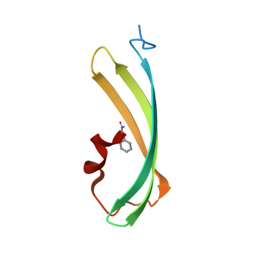
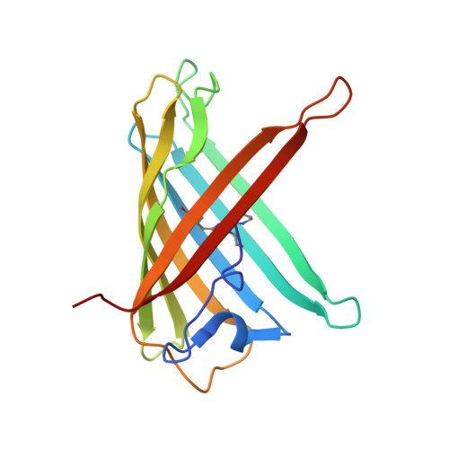
    Fluorescent proteins exhibit fluorescence quenching by specific transition metals, suggesting their potential as fluorescent protein-based metal biosensors. Each fluorescent protein exhibits unique spectroscopic properties and mechanisms for fluorescence quenching by metals. Therefore, the metal-induced fluorescence quenching analysis of various new fluorescent proteins would be important step towards the development of such fluorescent protein-based metal biosensors. Here, we first report the spectroscopic and structural analysis of the yellow fluorescent protein ZsYellow, following its metal-induced quenching. Spectroscopic analysis showed that ZsYellow exhibited a high degree of fluorescence quenching by Cu 2+ . During Cu 2+ -induced ZsYellow quenching, fluorescence emission was recovered by adding EDTA. The crystal structure of ZsYellow soaked in Cu 2+ solution was determined at a 2.6 Å resolution. The electron density map did not indicate the presence of Cu 2+ around the chromophore or the β-barrel surface, which resulted in fluorescence quenching without Cu 2+ binding to specific site in ZsYellow. Based on these results, we propose the fluorescence quenching to occur in a distance-dependent manner between the metal and the fluorescent protein, when these components get to a closer vicinity at higher metal concentrations. Our results provide useful insights for future development of fluorescent protein-based metal biosensors.


  • Organizational Affiliation
    • Division of Biotechnology, College of Life Sciences and Biotechnology, Korea University, Seoul 02841, Korea.

Macromolecule Content 

  • Total Structure Weight: 52.87 kDa 
  • Atom Count: 3,658 
  • Modeled Residue Count: 449 
  • Deposited Residue Count: 464 
  • Unique protein chains: 2

Macromolecules

Find similar proteins by:|  3D Structure
Entity ID: 1
MoleculeChains  Sequence LengthOrganismDetailsImage
GFP-like fluorescent chromoprotein FP538A [auth C],
C [auth D]
68Zoanthus sp.Mutation(s): 0 
UniProt
Find proteins for Q9U6Y4 (Zoanthus sp.)
Explore Q9U6Y4 
Go to UniProtKB:  Q9U6Y4
Entity Groups
Sequence Clusters30% Identity50% Identity70% Identity90% Identity95% Identity100% Identity
UniProt GroupQ9U6Y4
Sequence Annotations
Expand
Reference Sequence
Find similar proteins by:|  3D Structure
Entity ID: 2
MoleculeChains  Sequence LengthOrganismDetailsImage
GFP-like fluorescent chromoprotein FP538B [auth A],
D [auth B]
164Zoanthus sp.Mutation(s): 2 
UniProt
Find proteins for Q9U6Y4 (Zoanthus sp.)
Explore Q9U6Y4 
Go to UniProtKB:  Q9U6Y4
Entity Groups
Sequence Clusters30% Identity50% Identity70% Identity90% Identity95% Identity100% Identity
UniProt GroupQ9U6Y4
Sequence Annotations
Expand
Reference Sequence

Small Molecules

Modified Residues  2 Unique
IDChains TypeFormula2D DiagramParent
NFA
Query on NFA
A [auth C],
C [auth D]
L-PEPTIDE LINKINGC9 H12 N2 OPHE
CH7
Query on CH7
B [auth A],
D [auth B]
L-PEPTIDE LINKINGC17 H17 N3 O4LYS, TYR, GLY

Experimental Data & Validation

Experimental Data

  • Method: X-RAY DIFFRACTION
  • Resolution: 2.60 Å
  • R-Value Free:  0.215 (Depositor), 0.215 (DCC) 
  • R-Value Work:  0.203 (Depositor), 0.207 (DCC) 
  • R-Value Observed: 0.206 (Depositor) 
Space Group: P 21 2 21
Unit Cell:
Length ( Å )Angle ( ˚ )
a = 48.638α = 90
b = 72.929β = 90
c = 124.189γ = 90
Software Package:
Software NamePurpose
HKL-2000data scaling
REFMACrefinement
PDB_EXTRACTdata extraction
HKL-2000data reduction
MOLREPphasing

Structure Validation

View Full Validation Report


Entry History 

& Funding Information

Deposition Data

  • Released Date: 2020-01-22 
  • Deposition Author(s): Nam, K.H.

Funding OrganizationLocationGrant Number
National Research Foundation (NRF, Korea)Korea, Republic OfNRF-2017R1D1A1B03033087

Revision History  (Full details and data files)

  • Version 1.0: 2020-01-22
    Type: Initial release
  • Version 1.1: 2020-07-29
    Changes: Database references
  • Version 1.2: 2020-10-14
    Changes: Database references
  • Version 1.3: 2023-11-29
    Changes: Data collection, Database references, Refinement description
  • Version 2.0: 2026-03-18
    Changes: Polymer sequence, Structure summary