High-Performance Intensiometric Direct- and Inverse-Response Genetically Encoded Biosensors for Citrate.
Zhao, Y., Shen, Y., Wen, Y., Campbell, R.E.(2020) ACS Cent Sci 6: 1441-1450
- PubMed: 32875085 Search on PubMedSearch on PubMed Central
- DOI: https://doi.org/10.1021/acscentsci.0c00518
- Primary Citation Related Structures:
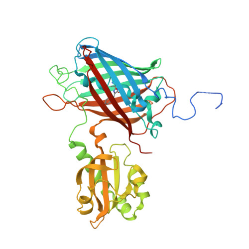
6LNP - PubMed Abstract:
Motivated by the growing recognition of citrate as a central metabolite in a variety of biological processes associated with healthy and diseased cellular states, we have developed a series of high-performance genetically encoded citrate biosensors suitable for imaging of citrate concentrations in mammalian cells. The design of these biosensors was guided by structural studies of the citrate-responsive sensor histidine kinase and took advantage of the same conformational changes proposed to propagate from the binding domain to the catalytic domain. Following extensive engineering based on a combination of structure guided mutagenesis and directed evolution, we produced an inverse-response biosensor (Δ F / F min ≈ 18) designated Citroff1 and a direct-response biosensor (Δ F / F min ≈ 9) designated Citron1. We report the X-ray crystal structure of Citron1 and demonstrate the utility of both biosensors for qualitative and quantitative imaging of steady-state and pharmacologically perturbed citrate concentrations in live cells.
- Department of Chemistry, University of Alberta, Edmonton, Alberta T6G 2G2, Canada.
Organizational Affiliation: