6JIL

Crystal structure of D-cycloserine synthetase DcsG


Experimental Data Snapshot

  • Method: X-RAY DIFFRACTION
  • Resolution: 2.32 Å
  • R-Value Free: 0.251 
  • R-Value Work: 0.195 
  • R-Value Observed: 0.195 

wwPDB Validation   3D Report Full Report


Ligand Structure Quality Assessment 


This is version 1.1 of the entry. See complete history


Literature

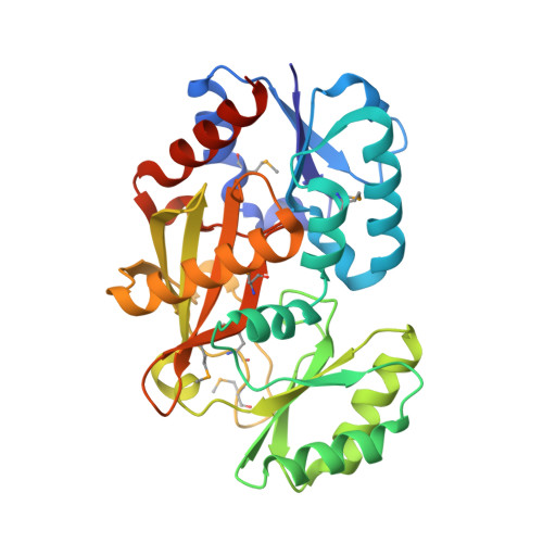
Cyclization mechanism catalyzed by an ATP-grasp enzyme essential for d-cycloserine biosynthesis.

Matoba, Y.Uda, N.Kudo, M.Sugiyama, M.

(2020) FEBS J 287: 2763-2778

  • DOI: https://doi.org/10.1111/febs.15163
  • Primary Citation of Related Structures:  
    6JIL

  • PubMed Abstract: 

    In the biosynthetic pathway of an antitubercular antibiotic d-cycloserine (d-CS), O-ureido-d-serine (d-OUS) is converted to d-CS. We have previously demonstrated that DcsG, classified into the ATP-grasp superfamily enzyme, catalyzes the ring formation to generate d-CS, which is accompanied by the cleavage of a bond in the urea moiety of d-OUS to remove a carbamoyl group. Although the general ATP-grasp enzymes catalyze an ATP-dependent ligation reaction between two substrates, DcsG catalyzes specifically the generation of an intramolecular covalent bond. In the present study, cyanate was found in the reaction mixture, suggesting that carbamoyl group is eliminated as an isocyanic acid during the reaction. By the crystallographic and mutational investigations of DcsG, we anticipate the residues necessary for the binding of d-OUS. An acylphosphate intermediate must be bound at the narrow pocket of DcsG in a folded conformation, inducing the bond cleavage and the new bond formation to generate cyanate and d-CS, respectively. DATABASE: Structural data are available in Protein Data Bank database under the accession number 6JIL.


  • Organizational Affiliation

    Faculty of Pharmacy, Yasuda Women's University, Hiroshima, Japan.


Macromolecules
Find similar proteins by:  (by identity cutoff)  |  3D Structure
Entity ID: 1
MoleculeChains Sequence LengthOrganismDetailsImage
Cycloserine biosynthesis protein DcsG
A, B, C, D
307Streptomyces lavendulaeMutation(s): 0 
Gene Names: dcsG
EC: 6.3.3.5
UniProt
Find proteins for D2Z030 (Streptomyces lavendulae)
Explore D2Z030 
Go to UniProtKB:  D2Z030
Entity Groups  
Sequence Clusters30% Identity50% Identity70% Identity90% Identity95% Identity100% Identity
UniProt GroupD2Z030
Sequence Annotations
Expand
  • Reference Sequence
Small Molecules
Ligands 3 Unique
IDChains Name / Formula / InChI Key2D Diagram3D Interactions
ADP
Query on ADP

Download Ideal Coordinates CCD File 
G [auth A],
M [auth B],
S [auth C],
X [auth D]
ADENOSINE-5'-DIPHOSPHATE
C10 H15 N5 O10 P2
XTWYTFMLZFPYCI-KQYNXXCUSA-N
TLA
Query on TLA

Download Ideal Coordinates CCD File 
E [auth A]
F [auth A]
J [auth B]
K [auth B]
L [auth B]
E [auth A],
F [auth A],
J [auth B],
K [auth B],
L [auth B],
P [auth C],
Q [auth C],
R [auth C],
V [auth D],
W [auth D]
L(+)-TARTARIC ACID
C4 H6 O6
FEWJPZIEWOKRBE-JCYAYHJZSA-N
MG
Query on MG

Download Ideal Coordinates CCD File 
AA [auth D]
H [auth A]
I [auth A]
N [auth B]
O [auth B]
AA [auth D],
H [auth A],
I [auth A],
N [auth B],
O [auth B],
T [auth C],
U [auth C],
Y [auth D],
Z [auth D]
MAGNESIUM ION
Mg
JLVVSXFLKOJNIY-UHFFFAOYSA-N
Modified Residues  1 Unique
IDChains TypeFormula2D DiagramParent
MSE
Query on MSE
A, B, C, D
L-PEPTIDE LINKINGC5 H11 N O2 SeMET
Experimental Data & Validation

Experimental Data

  • Method: X-RAY DIFFRACTION
  • Resolution: 2.32 Å
  • R-Value Free: 0.251 
  • R-Value Work: 0.195 
  • R-Value Observed: 0.195 
  • Space Group: P 1 21 1
Unit Cell:
Length ( Å )Angle ( ˚ )
a = 56.45α = 90
b = 120.73β = 101.01
c = 102.81γ = 90
Software Package:
Software NamePurpose
CNSrefinement
HKL-2000data reduction
HKL-2000data scaling
SHARPphasing

Structure Validation

View Full Validation Report


Ligand Structure Quality Assessment 


Entry History & Funding Information

Deposition Data


Funding OrganizationLocationGrant Number
Japan Society for the Promotion of ScienceJapan15K07997

Revision History  (Full details and data files)

  • Version 1.0: 2019-12-18
    Type: Initial release
  • Version 1.1: 2020-07-22
    Changes: Database references