6BXJ | pdb_00006bxj

Structure of a single-chain beta3 integrin


Experimental Data Snapshot

  • Method: X-RAY DIFFRACTION
  • Resolution: 2.09 Å
  • R-Value Free: 
    0.234 (Depositor), 0.234 (DCC) 
  • R-Value Work: 
    0.186 (Depositor), 0.187 (DCC) 
  • R-Value Observed: 
    0.189 (Depositor) 

wwPDB Validation   3D Report Full Report


Ligand Structure Quality Assessment 


This is version 1.5 of the entry. See complete history


Literature

Autonomous conformational regulation of beta3integrin and the conformation-dependent property of HPA-1a alloantibodies.

Thinn, A.M.M.Wang, Z.Zhou, D.Zhao, Y.Curtis, B.R.Zhu, J.

(2018) Proc Natl Acad Sci U S A 115: E9105-E9114

  • DOI: https://doi.org/10.1073/pnas.1806205115
  • Primary Citation of Related Structures:  
    6BXJ

  • PubMed Abstract: 

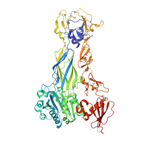
    Integrin α/β heterodimer adopts a compact bent conformation in the resting state, and upon activation undergoes a large-scale conformational rearrangement. During the inside-out activation, signals impinging on the cytoplasmic tail of β subunit induce the α/β separation at the transmembrane and cytoplasmic domains, leading to the extended conformation of the ectodomain with the separated leg and the opening headpiece that is required for the high-affinity ligand binding. It remains enigmatic which integrin subunit drives the bent-to-extended conformational rearrangement in the inside-out activation. The β 3 integrins, including α IIb β 3 and α V β 3 , are the prototypes for understanding integrin structural regulation. The Leu33Pro polymorphism located at the β 3 PSI domain defines the human platelet-specific alloantigen (HPA) 1a/b, which provokes the alloimmune response leading to clinically important bleeding disorders. Some, but not all, anti-HPA-1a alloantibodies can distinguish the α IIb β 3 from α V β 3 and affect their functions with unknown mechanisms. Here we designed a single-chain β 3 subunit that mimics a separation of α/β heterodimer on inside-out activation. Our crystallographic and functional studies show that the single-chain β 3 integrin folds into a bent conformation in solution but spontaneously extends on the cell surface. This demonstrates that the β 3 subunit autonomously drives the membrane-dependent conformational rearrangement during integrin activation. Using the single-chain β 3 integrin, we identified the conformation-dependent property of anti-HPA-1a alloantibodies, which enables them to differently recognize the β 3 in the bent state vs. the extended state and in the complex with α IIb vs. α V This study provides deeper understandings of integrin conformational activation on the cell surface.


  • Organizational Affiliation

    Blood Research Institute, BloodCenter of Wisconsin, Part of Versiti, Milwaukee, WI 53226.


Macromolecules
Find similar proteins by:  (by identity cutoff)  |  3D Structure
Entity ID: 1
MoleculeChains Sequence LengthOrganismDetailsImage
Chimera protein of Integrin beta-3 and Integrin alpha-L625Homo sapiensMutation(s): 0 
Gene Names: ITGB3GP3AITGALCD11A
UniProt & NIH Common Fund Data Resources
Find proteins for P05106 (Homo sapiens)
Explore P05106 
Go to UniProtKB:  P05106
PHAROS:  P05106
GTEx:  ENSG00000259207 
Find proteins for P20701 (Homo sapiens)
Explore P20701 
Go to UniProtKB:  P20701
PHAROS:  P20701
GTEx:  ENSG00000005844 
Entity Groups  
Sequence Clusters30% Identity50% Identity70% Identity90% Identity95% Identity100% Identity
UniProt GroupsP20701P05106
Glycosylation
Glycosylation Sites: 4Go to GlyGen: P20701-1P05106-1
Sequence Annotations
Expand
  • Reference Sequence
Experimental Data & Validation

Experimental Data

  • Method: X-RAY DIFFRACTION
  • Resolution: 2.09 Å
  • R-Value Free:  0.234 (Depositor), 0.234 (DCC) 
  • R-Value Work:  0.186 (Depositor), 0.187 (DCC) 
  • R-Value Observed: 0.189 (Depositor) 
Space Group: P 1 21 1
Unit Cell:
Length ( Å )Angle ( ˚ )
a = 55.46α = 90
b = 125.29β = 112.95
c = 70.02γ = 90
Software Package:
Software NamePurpose
PHENIXrefinement
SCALAdata scaling
PDB_EXTRACTdata extraction
TRUNCATEdata reduction
PHASERphasing

Structure Validation

View Full Validation Report


Ligand Structure Quality Assessment 


Entry History & Funding Information

Deposition Data


Funding OrganizationLocationGrant Number
National Institutes of Health/National Heart, Lung, and Blood Institute (NIH/NHLBI)United StatesHL131836
National Institutes of Health/National Heart, Lung, and Blood Institute (NIH/NHLBI)United StatesHL122985

Revision History  (Full details and data files)

  • Version 1.0: 2018-10-03
    Type: Initial release
  • Version 1.1: 2018-10-10
    Changes: Data collection, Database references, Structure summary
  • Version 1.2: 2019-02-20
    Changes: Author supporting evidence, Data collection, Structure summary
  • Version 1.3: 2019-12-04
    Changes: Author supporting evidence
  • Version 1.4: 2020-07-29
    Type: Remediation
    Reason: Carbohydrate remediation
    Changes: Data collection, Derived calculations, Structure summary
  • Version 1.5: 2024-10-09
    Changes: Data collection, Database references, Structure summary