5UB5

human POGLUT1 in complex with human Notch1 EGF12 S458T mutant and UDP


Experimental Data Snapshot

  • Method: X-RAY DIFFRACTION
  • Resolution: 2.09 Å
  • R-Value Free: 0.193 
  • R-Value Work: 0.153 
  • R-Value Observed: 0.155 

wwPDB Validation   3D Report Full Report


Ligand Structure Quality Assessment 


This is version 1.4 of the entry. See complete history


Literature

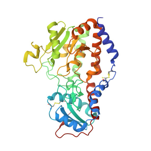
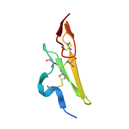
Structural basis of Notch O-glucosylation and O-xylosylation by mammalian protein-O-glucosyltransferase 1 (POGLUT1).

Li, Z.Fischer, M.Satkunarajah, M.Zhou, D.Withers, S.G.Rini, J.M.

(2017) Nat Commun 8: 185-185

  • DOI: https://doi.org/10.1038/s41467-017-00255-7
  • Primary Citation of Related Structures:  
    5L0R, 5L0S, 5L0T, 5L0U, 5L0V, 5UB5

  • PubMed Abstract: 

    Protein O-glucosyltransferase 1/Rumi-mediated glucosylation of Notch epidermal growth factor-like (EGF-like) domains plays an important role in Notch signaling. Protein O-glucosyltransferase 1 shows specificity for folded EGF-like domains, it can only glycosylate serine residues in the C 1 XSXPC 2 motif, and it possesses an uncommon dual donor substrate specificity. Using several EGF-like domains and donor substrate analogs, we have determined the structures of human Protein O-glucosyltransferase 1 substrate/product complexes that provide mechanistic insight into the basis for these properties. Notably, we show that Protein O-glucosyltransferase 1's requirement for folded EGF-like domains also leads to its serine specificity and that two distinct local conformational states are likely responsible for its ability to transfer both glucose and xylose. We also show that Protein O-glucosyltransferase 1 possesses the potential to xylosylate a much broader range of EGF-like domain substrates than was previously thought. Finally, we show that Protein O-glucosyltransferase 1 has co-evolved with EGF-like domains of the type found in Notch.POGLUT1 is a protein-O-glucosyltransferase that transfers glucose and xylose to the EGF-like domains of Notch and other signaling receptors. Here the authors report the structure of human POGLUT1 in complexes with 3 different EGF-like domains and donor substrates and shed light on the enzyme's substrate specificity and catalytic mechanism.


  • Organizational Affiliation

    Department of Biochemistry, University of Toronto, Toronto, ON, Canada, M5S 1A8.


Macromolecules
Find similar proteins by:  (by identity cutoff)  |  3D Structure
Entity ID: 1
MoleculeChains Sequence LengthOrganismDetailsImage
Protein O-glucosyltransferase 1357Homo sapiensMutation(s): 0 
Gene Names: POGLUT1C3orf9CLP46KTELC1MDSRPMDS010UNQ490/PRO1006
EC: 2.4.1 (PDB Primary Data), 2.4.2.26 (PDB Primary Data)
UniProt & NIH Common Fund Data Resources
Find proteins for Q8NBL1 (Homo sapiens)
Explore Q8NBL1 
Go to UniProtKB:  Q8NBL1
PHAROS:  Q8NBL1
GTEx:  ENSG00000163389 
Entity Groups  
Sequence Clusters30% Identity50% Identity70% Identity90% Identity95% Identity100% Identity
UniProt GroupQ8NBL1
Sequence Annotations
Expand
  • Reference Sequence
Find similar proteins by:  (by identity cutoff)  |  3D Structure
Entity ID: 2
MoleculeChains Sequence LengthOrganismDetailsImage
Neurogenic locus notch homolog protein 142Homo sapiensMutation(s): 1 
Gene Names: NOTCH1TAN1
UniProt & NIH Common Fund Data Resources
Find proteins for P46531 (Homo sapiens)
Explore P46531 
Go to UniProtKB:  P46531
PHAROS:  P46531
GTEx:  ENSG00000148400 
Entity Groups  
Sequence Clusters30% Identity50% Identity70% Identity90% Identity95% Identity100% Identity
UniProt GroupP46531
Sequence Annotations
Expand
  • Reference Sequence
Experimental Data & Validation

Experimental Data

  • Method: X-RAY DIFFRACTION
  • Resolution: 2.09 Å
  • R-Value Free: 0.193 
  • R-Value Work: 0.153 
  • R-Value Observed: 0.155 
  • Space Group: P 21 21 21
Unit Cell:
Length ( Å )Angle ( ˚ )
a = 70.951α = 90
b = 74.072β = 90
c = 83.583γ = 90
Software Package:
Software NamePurpose
PHENIXrefinement
SAINTdata scaling
PDB_EXTRACTdata extraction
SAINTdata reduction
PHASERphasing

Structure Validation

View Full Validation Report


Ligand Structure Quality Assessment 


Entry History 

Deposition Data

Revision History  (Full details and data files)

  • Version 1.0: 2017-08-09
    Type: Initial release
  • Version 1.1: 2017-08-30
    Changes: Database references
  • Version 1.2: 2018-04-04
    Changes: Data collection, Source and taxonomy
  • Version 1.3: 2020-07-29
    Type: Remediation
    Reason: Carbohydrate remediation
    Changes: Data collection, Derived calculations, Structure summary
  • Version 1.4: 2023-10-04
    Changes: Data collection, Database references, Refinement description, Structure summary