5OV7 | pdb_00005ov7

tubulin - rigosertib complex


Experimental Data Snapshot

  • Method: X-RAY DIFFRACTION
  • Resolution: 2.40 Å
  • R-Value Free: 
    0.235 (Depositor), 0.237 (DCC) 
  • R-Value Work: 
    0.190 (Depositor), 0.193 (DCC) 
  • R-Value Observed: 
    0.192 (Depositor) 

Starting Model: experimental
View more details

wwPDB Validation 3D Report Full Report

Validation slider image for 5OV7

Ligand Structure Quality Assessment 


This is version 1.2 of the entry. See complete history

Literature

Combined CRISPRi/a-Based Chemical Genetic Screens Reveal that Rigosertib Is a Microtubule-Destabilizing Agent.

Jost, M.Chen, Y.Gilbert, L.A.Horlbeck, M.A.Krenning, L.Menchon, G.Rai, A.Cho, M.Y.Stern, J.J.Prota, A.E.Kampmann, M.Akhmanova, A.Steinmetz, M.O.Tanenbaum, M.E.Weissman, J.S.

(2017) Mol Cell 68: 210-223.e6

  • DOI: https://doi.org/10.1016/j.molcel.2017.09.012
  • Primary Citation Related Structures: 
    5OV7

  • PubMed Abstract: 

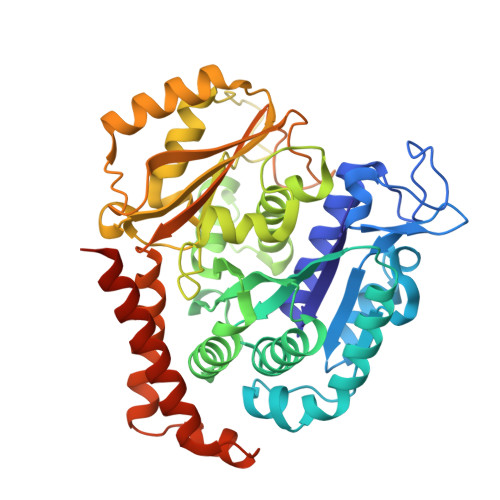
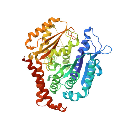
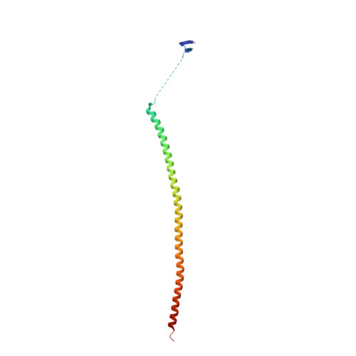
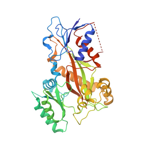
    Chemical libraries paired with phenotypic screens can now readily identify compounds with therapeutic potential. A central limitation to exploiting these compounds, however, has been in identifying their relevant cellular targets. Here, we present a two-tiered CRISPR-mediated chemical-genetic strategy for target identification: combined genome-wide knockdown and overexpression screening as well as focused, comparative chemical-genetic profiling. Application of these strategies to rigosertib, a drug in phase 3 clinical trials for high-risk myelodysplastic syndrome whose molecular target had remained controversial, pointed singularly to microtubules as rigosertib's target. We showed that rigosertib indeed directly binds to and destabilizes microtubules using cell biological, in vitro, and structural approaches. Finally, expression of tubulin with a structure-guided mutation in the rigosertib-binding pocket conferred resistance to rigosertib, establishing that rigosertib kills cancer cells by destabilizing microtubules. These results demonstrate the power of our chemical-genetic screening strategies for pinpointing the physiologically relevant targets of chemical agents.


  • Organizational Affiliation
    • Department of Cellular and Molecular Pharmacology, University of California, San Francisco, San Francisco, CA 94158, USA; Howard Hughes Medical Institute, University of California, San Francisco, San Francisco, CA 94158, USA; Center for RNA Systems Biology, University of California, San Francisco, San Francisco, CA 94158, USA; Department of Microbiology and Immunology, University of California, San Francisco, San Francisco, CA 94158, USA.

Macromolecule Content 

  • Total Structure Weight: 265.88 kDa 
  • Atom Count: 17,978 
  • Modeled Residue Count: 2,189 
  • Deposited Residue Count: 2,319 
  • Unique protein chains: 4

Macromolecules

Find similar proteins by:|  3D Structure
Entity ID: 1
MoleculeChains  Sequence LengthOrganismDetailsImage
Tubulin alpha-1B chain
A, C
451Bos taurusMutation(s): 0 
EC: 3.6.5
UniProt
Find proteins for P81947 (Bos taurus)
Explore P81947 
Go to UniProtKB:  P81947
Entity Groups
Sequence Clusters30% Identity50% Identity70% Identity90% Identity95% Identity100% Identity
UniProt GroupP81947
Sequence Annotations
Expand
Reference Sequence
Find similar proteins by:|  3D Structure
Entity ID: 2
MoleculeChains  Sequence LengthOrganismDetailsImage
Tubulin beta-2B chain
B, D
445Bos taurusMutation(s): 0 
UniProt
Find proteins for Q6B856 (Bos taurus)
Explore Q6B856 
Go to UniProtKB:  Q6B856
Entity Groups
Sequence Clusters30% Identity50% Identity70% Identity90% Identity95% Identity100% Identity
UniProt GroupQ6B856
Sequence Annotations
Expand
Reference Sequence
Find similar proteins by:|  3D Structure
Entity ID: 3
MoleculeChains  Sequence LengthOrganismDetailsImage
Stathmin-4143Rattus norvegicusMutation(s): 0 
Gene Names: Stmn4
UniProt
Find proteins for P63043 (Rattus norvegicus)
Explore P63043 
Go to UniProtKB:  P63043
Entity Groups
Sequence Clusters30% Identity50% Identity70% Identity90% Identity95% Identity100% Identity
UniProt GroupP63043
Sequence Annotations
Expand
Reference Sequence
Find similar proteins by:|  3D Structure
Entity ID: 4
MoleculeChains  Sequence LengthOrganismDetailsImage
Uncharacterized protein384Gallus gallusMutation(s): 0 
Gene Names: TTL

Small Molecules

Ligands 8 Unique
IDChains Name / Formula / InChI Key2D Diagram3D Interactions
GTP

Query on GTP


Download:Ideal Coordinates CCD File
G [auth A],
S [auth C]
GUANOSINE-5'-TRIPHOSPHATE
C10 H16 N5 O14 P3
XKMLYUALXHKNFT-UUOKFMHZSA-N
ACP

Query on ACP


Download:Ideal Coordinates CCD File
Y [auth F]PHOSPHOMETHYLPHOSPHONIC ACID ADENYLATE ESTER
C11 H18 N5 O12 P3
UFZTZBNSLXELAL-IOSLPCCCSA-N
6FS

Query on 6FS


Download:Ideal Coordinates CCD File
O [auth B],
X [auth D]
N-[2-methoxy-5-({[(E)-2-(2,4,6-trimethoxyphenyl)ethenyl]sulfonyl}methyl)phenyl]glycine
C21 H25 N O8 S
OWBFCJROIKNMGD-BQYQJAHWSA-N
GDP

Query on GDP


Download:Ideal Coordinates CCD File
L [auth B],
V [auth D]
GUANOSINE-5'-DIPHOSPHATE
C10 H15 N5 O11 P2
QGWNDRXFNXRZMB-UUOKFMHZSA-N
MES

Query on MES


Download:Ideal Coordinates CCD File
P [auth B]2-(N-MORPHOLINO)-ETHANESULFONIC ACID
C6 H13 N O4 S
SXGZJKUKBWWHRA-UHFFFAOYSA-N
GOL

Query on GOL


Download:Ideal Coordinates CCD File
J [auth A]
K [auth A]
N [auth B]
Q [auth B]
R [auth C]
J [auth A],
K [auth A],
N [auth B],
Q [auth B],
R [auth C],
U [auth C]
GLYCEROL
C3 H8 O3
PEDCQBHIVMGVHV-UHFFFAOYSA-N
CA

Query on CA


Download:Ideal Coordinates CCD File
I [auth A]CALCIUM ION
Ca
BHPQYMZQTOCNFJ-UHFFFAOYSA-N
MG

Query on MG


Download:Ideal Coordinates CCD File
H [auth A],
M [auth B],
T [auth C],
W [auth D],
Z [auth F]
MAGNESIUM ION
Mg
JLVVSXFLKOJNIY-UHFFFAOYSA-N

Experimental Data & Validation

Experimental Data

  • Method: X-RAY DIFFRACTION
  • Resolution: 2.40 Å
  • R-Value Free:  0.235 (Depositor), 0.237 (DCC) 
  • R-Value Work:  0.190 (Depositor), 0.193 (DCC) 
  • R-Value Observed: 0.192 (Depositor) 
Space Group: P 21 21 21
Unit Cell:
Length ( Å )Angle ( ˚ )
a = 104.732α = 90
b = 156.765β = 90
c = 182.629γ = 90
Software Package:
Software NamePurpose
PHENIXrefinement
XDSdata reduction
XDSdata scaling
PHENIXphasing

Structure Validation

View Full Validation Report


Ligand Structure Quality Assessment 


Entry History 

Deposition Data

Revision History  (Full details and data files)

  • Version 1.0: 2017-10-11
    Type: Initial release
  • Version 1.1: 2017-10-18
    Changes: Database references
  • Version 1.2: 2024-01-17
    Changes: Data collection, Database references, Derived calculations, Refinement description