5JJK

Rho transcription termination factor bound to rA7 and 6 ADP-BeF3 molecules


Experimental Data Snapshot

  • Method: X-RAY DIFFRACTION
  • Resolution: 3.15 Å
  • R-Value Free: 0.265 
  • R-Value Work: 0.233 
  • R-Value Observed: 0.235 

wwPDB Validation   3D Report Full Report


Ligand Structure Quality Assessment 


This is version 1.6 of the entry. See complete history


Literature

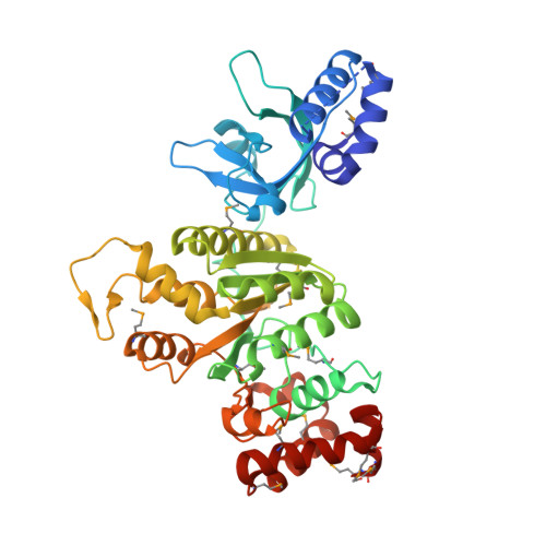
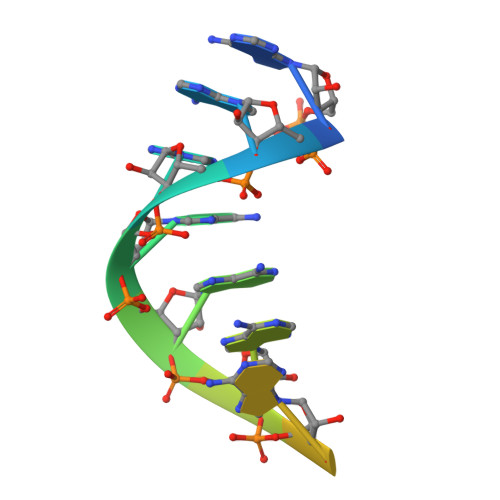
Molecular mechanisms of substrate-controlled ring dynamics and substepping in a nucleic acid-dependent hexameric motor.

Thomsen, N.D.Lawson, M.R.Witkowsky, L.B.Qu, S.Berger, J.M.

(2016) Proc Natl Acad Sci U S A 113: E7691-E7700

  • DOI: https://doi.org/10.1073/pnas.1616745113
  • Primary Citation of Related Structures:  
    5JJI, 5JJK, 5JJL

  • PubMed Abstract: 

    Ring-shaped hexameric helicases and translocases support essential DNA-, RNA-, and protein-dependent transactions in all cells and many viruses. How such systems coordinate ATPase activity between multiple subunits to power conformational changes that drive the engagement and movement of client substrates is a fundamental question. Using the Escherichia coli Rho transcription termination factor as a model system, we have used solution and crystallographic structural methods to delineate the range of conformational changes that accompany distinct substrate and nucleotide cofactor binding events. Small-angle X-ray scattering data show that Rho preferentially adopts an open-ring state in solution and that RNA and ATP are both required to cooperatively promote ring closure. Multiple closed-ring structures with different RNA substrates and nucleotide occupancies capture distinct catalytic intermediates accessed during translocation. Our data reveal how RNA-induced ring closure templates a sequential ATP-hydrolysis mechanism, provide a molecular rationale for how the Rho ATPase domains distinguishes between distinct RNA sequences, and establish structural snapshots of substepping events in a hexameric helicase/translocase.


  • Organizational Affiliation

    Department of Molecular and Cell Biology, University of California, Berkeley, CA 94720.


Macromolecules

Find similar proteins by:  (by identity cutoff)  |  3D Structure
Entity ID: 1
MoleculeChains Sequence LengthOrganismDetailsImage
Transcription termination factor Rho
A, B, C, D, E
A, B, C, D, E, F
420Escherichia coli O157:H7Mutation(s): 0 
Gene Names: rhoZ5293ECs4716
EC: 3.6.4
UniProt
Find proteins for P0AG30 (Escherichia coli (strain K12))
Explore P0AG30 
Go to UniProtKB:  P0AG30
Entity Groups  
Sequence Clusters30% Identity50% Identity70% Identity90% Identity95% Identity100% Identity
UniProt GroupP0AG30
Sequence Annotations
Expand
  • Reference Sequence

Find similar nucleic acids by:  Sequence   |   3D Structure  

Entity ID: 2
MoleculeChains LengthOrganismImage
rA12: RNA (5'-R(P*AP*AP*AP*AP*AP*AP*A)-3')12synthetic construct
Sequence Annotations
Expand
  • Reference Sequence
Small Molecules
Ligands 3 Unique
IDChains Name / Formula / InChI Key2D Diagram3D Interactions
ADP
Query on ADP

Download Ideal Coordinates CCD File 
H [auth A]
K [auth B]
N [auth C]
Q [auth D]
T [auth E]
H [auth A],
K [auth B],
N [auth C],
Q [auth D],
T [auth E],
W [auth F]
ADENOSINE-5'-DIPHOSPHATE
C10 H15 N5 O10 P2
XTWYTFMLZFPYCI-KQYNXXCUSA-N
BEF
Query on BEF

Download Ideal Coordinates CCD File 
J [auth A]
M [auth B]
P [auth C]
S [auth D]
V [auth E]
J [auth A],
M [auth B],
P [auth C],
S [auth D],
V [auth E],
Y [auth F]
BERYLLIUM TRIFLUORIDE ION
Be F3
OGIAHMCCNXDTIE-UHFFFAOYSA-K
MG
Query on MG

Download Ideal Coordinates CCD File 
I [auth A]
L [auth B]
O [auth C]
R [auth D]
U [auth E]
I [auth A],
L [auth B],
O [auth C],
R [auth D],
U [auth E],
X [auth F]
MAGNESIUM ION
Mg
JLVVSXFLKOJNIY-UHFFFAOYSA-N
Modified Residues  1 Unique
IDChains TypeFormula2D DiagramParent
MSE
Query on MSE
A, B, C, D, E
A, B, C, D, E, F
L-PEPTIDE LINKINGC5 H11 N O2 SeMET
Experimental Data & Validation

Experimental Data

  • Method: X-RAY DIFFRACTION
  • Resolution: 3.15 Å
  • R-Value Free: 0.265 
  • R-Value Work: 0.233 
  • R-Value Observed: 0.235 
  • Space Group: P 1 21 1
Unit Cell:
Length ( Å )Angle ( ˚ )
a = 68.76α = 90
b = 199.21β = 104.83
c = 112.2γ = 90
Software Package:
Software NamePurpose
HKL-2000data scaling
PHENIXrefinement
PDB_EXTRACTdata extraction
HKL-2000data reduction

Structure Validation

View Full Validation Report


Ligand Structure Quality Assessment 


Entry History & Funding Information

Deposition Data


Funding OrganizationLocationGrant Number
National Institutes of Health/National Institute of General Medical Sciences (NIH/NIGMS)United StatesGM071747
G. Harold and Leila Y. Mathers FoundationUnited States--

Revision History  (Full details and data files)

  • Version 1.0: 2016-11-16
    Type: Initial release
  • Version 1.1: 2016-12-21
    Changes: Structure summary
  • Version 1.2: 2016-12-28
    Changes: Database references
  • Version 1.3: 2017-09-13
    Changes: Author supporting evidence
  • Version 1.4: 2019-12-25
    Changes: Author supporting evidence
  • Version 1.5: 2023-09-27
    Changes: Data collection, Database references, Derived calculations, Refinement description
  • Version 1.6: 2023-11-15
    Changes: Data collection