4Y5W

Transcription factor-DNA complex


Experimental Data Snapshot

  • Method: X-RAY DIFFRACTION
  • Resolution: 3.10 Å
  • R-Value Free: 0.278 
  • R-Value Work: 0.250 
  • R-Value Observed: 0.251 

wwPDB Validation   3D Report Full Report


This is version 1.6 of the entry. See complete history


Literature

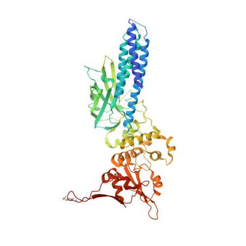
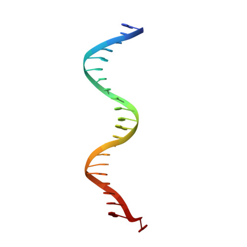
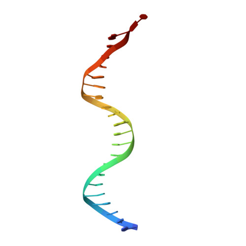
Structural basis for DNA recognition by STAT6

Li, J.Rodriguez, J.P.Niu, F.Pu, M.Wang, J.Hung, L.W.Shao, Q.Zhu, Y.Ding, W.Liu, Y.Da, Y.Yao, Z.Yang, J.Zhao, Y.Wei, G.H.Cheng, G.Liu, Z.J.Ouyang, S.

(2016) Proc Natl Acad Sci U S A 113: 13015-13020

  • DOI: https://doi.org/10.1073/pnas.1611228113
  • Primary Citation of Related Structures:  
    4Y5U, 4Y5W, 5D39

  • PubMed Abstract: 

    STAT6 participates in classical IL-4/IL-13 signaling and stimulator of interferon genes-mediated antiviral innate immune responses. Aberrations in STAT6-mediated signaling are linked to development of asthma and diseases of the immune system. In addition, STAT6 remains constitutively active in multiple types of cancer. Therefore, targeting STAT6 is an attractive proposition for treating related diseases. Although a lot is known about the role of STAT6 in transcriptional regulation, molecular details on how STAT6 recognizes and binds specific segments of DNA to exert its function are not clearly understood. Here, we report the crystal structures of a homodimer of phosphorylated STAT6 core fragment (STAT6 CF ) alone and bound with the N3 and N4 DNA binding site. Analysis of the structures reveals that STAT6 undergoes a dramatic conformational change on DNA binding, which was further validated by performing molecular dynamics simulation studies and small angle X-ray scattering analysis. Our data show that a larger angle at the intersection where the two protomers of STAT meet and the presence of a unique residue, H415, in the DNA-binding domain play important roles in discrimination of the N4 site DNA from the N3 site by STAT6. H415N mutation of STAT6 CF decreased affinity of the protein for the N4 site DNA, but increased its affinity for N3 site DNA, both in vitro and in vivo. Results of our structure-function studies on STAT6 shed light on mechanism of DNA recognition by STATs in general and explain the reasons underlying STAT6's preference for N4 site DNA over N3.


  • Organizational Affiliation

    National Laboratory of Biomacromolecules, Institute of Biophysics, Chinese Academy of Sciences, Beijing 100101, China.


Macromolecules

Find similar proteins by:  (by identity cutoff)  |  3D Structure
Entity ID: 1
MoleculeChains Sequence LengthOrganismDetailsImage
Signal transducer and activator of transcription 6A,
B,
C [auth D],
H [auth C]
549Homo sapiensMutation(s): 0 
Gene Names: STAT6
UniProt & NIH Common Fund Data Resources
Find proteins for P42226 (Homo sapiens)
Explore P42226 
Go to UniProtKB:  P42226
PHAROS:  P42226
GTEx:  ENSG00000166888 
Entity Groups  
Sequence Clusters30% Identity50% Identity70% Identity90% Identity95% Identity100% Identity
UniProt GroupP42226
Sequence Annotations
Expand
  • Reference Sequence

Find similar nucleic acids by:  Sequence   |   3D Structure  

Entity ID: 2
MoleculeChains LengthOrganismImage
DNA (5'-D(P*AP*TP*GP*GP*AP*TP*TP*TP*CP*CP*TP*AP*GP*GP*AP*AP*GP*AP*CP*AP*A)-3')D [auth E],
F [auth M]
22Homo sapiens
Sequence Annotations
Expand
  • Reference Sequence

Find similar nucleic acids by:  Sequence   |   3D Structure  

Entity ID: 3
MoleculeChains LengthOrganismImage
DNA (5'-D(P*TP*TP*GP*TP*CP*TP*TP*CP*CP*TP*AP*GP*GP*AP*AP*AP*TP*CP*CP*AP*T)-3')E [auth F],
G [auth N]
22Homo sapiens
Sequence Annotations
Expand
  • Reference Sequence
Small Molecules
Modified Residues  1 Unique
IDChains TypeFormula2D DiagramParent
PTR
Query on PTR
A,
B,
C [auth D],
H [auth C]
L-PEPTIDE LINKINGC9 H12 N O6 PTYR
Experimental Data & Validation

Experimental Data

  • Method: X-RAY DIFFRACTION
  • Resolution: 3.10 Å
  • R-Value Free: 0.278 
  • R-Value Work: 0.250 
  • R-Value Observed: 0.251 
  • Space Group: P 1
Unit Cell:
Length ( Å )Angle ( ˚ )
a = 68.298α = 99.85
b = 94.241β = 101.73
c = 147.785γ = 89.93
Software Package:
Software NamePurpose
PHENIXrefinement
PHENIXmodel building
PHASERphasing
HKL-2000data collection
HKL-2000data reduction
HKL-2000data scaling

Structure Validation

View Full Validation Report


Entry History 

Deposition Data

Revision History  (Full details and data files)

  • Version 1.0: 2016-02-17
    Type: Initial release
  • Version 1.1: 2016-03-02
    Changes: Data collection
  • Version 1.2: 2016-11-02
    Changes: Database references
  • Version 1.3: 2016-11-16
    Changes: Database references
  • Version 1.4: 2016-11-30
    Changes: Database references
  • Version 1.5: 2023-11-08
    Changes: Data collection, Database references, Refinement description
  • Version 1.6: 2023-11-15
    Changes: Data collection