4MQW | pdb_00004mqw

Structure of follicle-stimulating hormone in complex with the entire ectodomain of its receptor (P31)


Experimental Data Snapshot

  • Method: X-RAY DIFFRACTION
  • Resolution: 2.90 Å
  • R-Value Free: 
    0.237 (Depositor), 0.231 (DCC) 
  • R-Value Work: 
    0.174 (Depositor) 
  • R-Value Observed: 
    0.177 (Depositor) 

Starting Model: experimental
View more details

wwPDB Validation 3D Report Full Report

Validation slider image for 4MQW

Ligand Structure Quality Assessment 


This is version 1.7 of the entry. See complete history

Literature

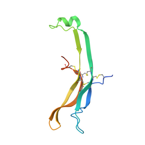
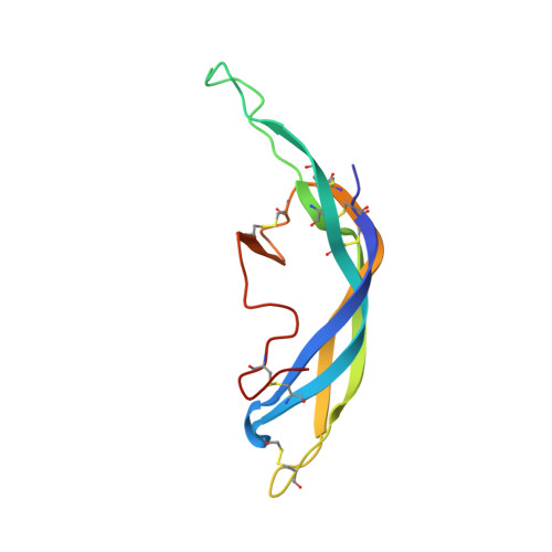
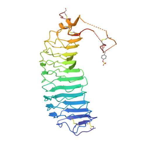
Evidence for Follicle-stimulating Hormone Receptor as a Functional Trimer.

Jiang, X.Fischer, D.Chen, X.McKenna, S.D.Liu, H.Sriraman, V.Yu, H.N.Goutopoulos, A.Arkinstall, S.He, X.

(2014) J Biological Chem 289: 14273-14282

  • DOI: https://doi.org/10.1074/jbc.M114.549592
  • Primary Citation Related Structures: 
    4MQW

  • PubMed Abstract: 

    Follicle-stimulating hormone receptor (FSHR), a G-protein coupled receptor, is an important drug target in the development of novel therapeutics for reproductive indications. The FSHR extracellular domains were observed in the crystal structure as a trimer, which enabled us to propose a novel model for the receptor activation mechanism. The model predicts that FSHR binds Asnα(52)-deglycosylated FSH at a 3-fold higher capacity than fully glycosylated FSH. It also predicts that, upon dissociation of the FSHR trimer into monomers, the binding of glycosylated FSH, but not deglycosylated FSH, would increase 3-fold, and that the dissociated monomers would in turn enhance FSHR binding and signaling activities by 3-fold. This study presents evidence confirming these predictions and provides crystallographic and mutagenesis data supporting the proposed model. The model also provides a mechanistic explanation to the agonist and antagonist activities of thyroid-stimulating hormone receptor autoantibodies. We conclude that FSHR exists as a functional trimer.


  • Organizational Affiliation
    • From the EMD Serono Research and Development Institute, Billerica, Massachusetts 01821 and xuliang.jiang@emdserono.com.

Macromolecule Content 

  • Total Structure Weight: 199.82 kDa 
  • Atom Count: 11,798 
  • Modeled Residue Count: 1,453 
  • Deposited Residue Count: 1,722 
  • Unique protein chains: 3

Macromolecules

Find similar proteins by:|  3D Structure
Entity ID: 1
MoleculeChains  Sequence LengthOrganismDetailsImage
Glycoprotein hormones, alpha polypeptide
A, D, G
102Homo sapiensMutation(s): 0 
Gene Names: CGA
UniProt & NIH Common Fund Data Resources
Find proteins for P01215 (Homo sapiens)
Explore P01215 
Go to UniProtKB:  P01215
PHAROS:  P01215
GTEx:  ENSG00000135346 
Entity Groups
Sequence Clusters30% Identity50% Identity70% Identity90% Identity95% Identity100% Identity
UniProt GroupP01215
Glycosylation
Glycosylation Sites: 2Go to GlyGen: P01215-1
Sequence Annotations
Expand
Reference Sequence
Find similar proteins by:|  3D Structure
Entity ID: 2
MoleculeChains  Sequence LengthOrganismDetailsImage
Follitropin subunit beta
B, E, H
111Homo sapiensMutation(s): 0 
Gene Names: FSHB
UniProt & NIH Common Fund Data Resources
Find proteins for P01225 (Homo sapiens)
Explore P01225 
Go to UniProtKB:  P01225
PHAROS:  P01225
GTEx:  ENSG00000131808 
Entity Groups
Sequence Clusters30% Identity50% Identity70% Identity90% Identity95% Identity100% Identity
UniProt GroupP01225
Glycosylation
Glycosylation Sites: 2Go to GlyGen: P01225-1
Sequence Annotations
Expand
Reference Sequence
Find similar proteins by:|  3D Structure
Entity ID: 3
MoleculeChains  Sequence LengthOrganismDetailsImage
Follicle-stimulating hormone receptorC [auth X],
F [auth Y],
I [auth Z]
361Homo sapiensMutation(s): 1 
Gene Names: FSHRLGR1
UniProt & NIH Common Fund Data Resources
Find proteins for P23945 (Homo sapiens)
Explore P23945 
Go to UniProtKB:  P23945
PHAROS:  P23945
GTEx:  ENSG00000170820 
Entity Groups
Sequence Clusters30% Identity50% Identity70% Identity90% Identity95% Identity100% Identity
UniProt GroupP23945
Glycosylation
Glycosylation Sites: 1Go to GlyGen: P23945-1
Sequence Annotations
Expand
Reference Sequence

Small Molecules

Ligands 3 Unique
IDChains Name / Formula / InChI Key2D Diagram3D Interactions
JEF

Query on JEF


Download:Ideal Coordinates CCD File
CA [auth Z]O-(O-(2-AMINOPROPYL)-O'-(2-METHOXYETHYL)POLYPROPYLENE GLYCOL 500)
C30 H63 N O10
ICCXIDTYQFYPNV-RUMGZKRTSA-N
NAG

Query on NAG


Download:Ideal Coordinates CCD File
AA [auth H]
BA [auth H]
DA [auth Z]
J [auth A]
K [auth A]
AA [auth H],
BA [auth H],
DA [auth Z],
J [auth A],
K [auth A],
L [auth B],
M [auth B],
R [auth X],
S [auth D],
T [auth D],
U [auth E],
V [auth E],
X [auth Y],
Y [auth G],
Z [auth G]
2-acetamido-2-deoxy-beta-D-glucopyranose
C8 H15 N O6
OVRNDRQMDRJTHS-FMDGEEDCSA-N
EDO

Query on EDO


Download:Ideal Coordinates CCD File
N [auth B],
O [auth B],
P [auth X],
Q [auth X],
W [auth Y]
1,2-ETHANEDIOL
C2 H6 O2
LYCAIKOWRPUZTN-UHFFFAOYSA-N
Modified Residues  1 Unique
IDChains TypeFormula2D DiagramParent
TYS
Query on TYS
C [auth X],
F [auth Y],
I [auth Z]
L-PEPTIDE LINKINGC9 H11 N O6 STYR

Experimental Data & Validation

Experimental Data

  • Method: X-RAY DIFFRACTION
  • Resolution: 2.90 Å
  • R-Value Free:  0.237 (Depositor), 0.231 (DCC) 
  • R-Value Work:  0.174 (Depositor) 
  • R-Value Observed: 0.177 (Depositor) 
Space Group: P 31
Unit Cell:
Length ( Å )Angle ( ˚ )
a = 95.9α = 90
b = 95.9β = 90
c = 204.281γ = 120
Software Package:
Software NamePurpose
HKL-3000data collection
PHASESphasing
REFMACrefinement
HKL-3000data reduction
HKL-3000data scaling
BUSTERrefinement

Structure Validation

View Full Validation Report


Ligand Structure Quality Assessment 


Entry History 

Deposition Data

Revision History  (Full details and data files)

  • Version 1.0: 2014-04-09
    Type: Initial release
  • Version 1.1: 2014-06-18
    Changes: Database references
  • Version 1.2: 2014-09-24
    Changes: Database references
  • Version 1.3: 2020-07-29
    Type: Remediation
    Reason: Carbohydrate remediation
    Changes: Data collection, Database references, Derived calculations, Structure summary
  • Version 1.4: 2023-09-20
    Changes: Data collection, Database references, Refinement description, Structure summary
  • Version 1.5: 2023-12-06
    Changes: Data collection
  • Version 1.6: 2024-01-10
    Changes: Derived calculations
  • Version 1.7: 2024-11-06
    Changes: Structure summary