4LOC

Structure of the carboxyl transferase domain from Rhizobium etli pyruvate carboxylase with oxamate and biotin


Experimental Data Snapshot

  • Method: X-RAY DIFFRACTION
  • Resolution: 2.26 Å
  • R-Value Free: 0.216 
  • R-Value Work: 0.176 
  • R-Value Observed: 0.178 

wwPDB Validation   3D Report Full Report


Ligand Structure Quality Assessment 


This is version 1.5 of the entry. See complete history


Literature

The role of biotin and oxamate in the carboxyltransferase reaction of pyruvate carboxylase.

Lietzan, A.D.Lin, Y.St.Maurice, M.

(2014) Arch Biochem Biophys 562C: 70-79

  • DOI: https://doi.org/10.1016/j.abb.2014.08.008
  • Primary Citation of Related Structures:  
    4LOC, 4M6V

  • PubMed Abstract: 

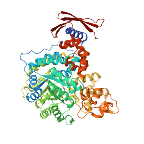
    Pyruvate carboxylase (PC) is a biotin-dependent enzyme that catalyzes the MgATP-dependent carboxylation of pyruvate to oxaloacetate, an important anaplerotic reaction in central metabolism. During catalysis, carboxybiotin is translocated to the carboxyltransferase domain where the carboxyl group is transferred to the acceptor substrate, pyruvate. Many studies on the carboxyltransferase domain of PC have demonstrated an enhanced oxaloacetate decarboxylation activity in the presence of oxamate and it has been shown that oxamate accepts a carboxyl group from carboxybiotin during oxaloacetate decarboxylation. The X-ray crystal structure of the carboxyltransferase domain from Rhizobium etli PC reveals that oxamate is positioned in the active site in an identical manner to the substrate, pyruvate, and kinetic data are consistent with the oxamate-stimulated decarboxylation of oxaloacetate proceeding through a simple ping-pong bi bi mechanism in the absence of the biotin carboxylase domain. Additionally, analysis of truncated PC enzymes indicates that the BCCP domain devoid of biotin does not contribute directly to the enzymatic reaction and conclusively demonstrates a biotin-independent oxaloacetate decarboxylation activity in PC. These findings advance the description of catalysis in PC and can be extended to the study of related biotin-dependent enzymes.


  • Organizational Affiliation

    Department of Biological Sciences, Marquette University, Milwaukee, WI 53201, USA.


Macromolecules
Find similar proteins by:  (by identity cutoff)  |  3D Structure
Entity ID: 1
MoleculeChains Sequence LengthOrganismDetailsImage
Pyruvate carboxylase
A, B, C, D
632Rhizobium etli CFN 42Mutation(s): 0 
Gene Names: pycRHE_CH04002
EC: 6.4.1.1
UniProt
Find proteins for Q2K340 (Rhizobium etli (strain CFN 42 / ATCC 51251))
Explore Q2K340 
Go to UniProtKB:  Q2K340
Entity Groups  
Sequence Clusters30% Identity50% Identity70% Identity90% Identity95% Identity100% Identity
UniProt GroupQ2K340
Sequence Annotations
Expand
  • Reference Sequence
Small Molecules
Ligands 6 Unique
IDChains Name / Formula / InChI Key2D Diagram3D Interactions
BTN
Query on BTN

Download Ideal Coordinates CCD File 
H [auth A],
M [auth B],
S [auth C],
X [auth D]
BIOTIN
C10 H16 N2 O3 S
YBJHBAHKTGYVGT-ZKWXMUAHSA-N
GOL
Query on GOL

Download Ideal Coordinates CCD File 
J [auth B],
Q [auth C]
GLYCEROL
C3 H8 O3
PEDCQBHIVMGVHV-UHFFFAOYSA-N
OXM
Query on OXM

Download Ideal Coordinates CCD File 
I [auth A],
N [auth B],
T [auth C],
Y [auth D]
OXAMIC ACID
C2 H3 N O3
SOWBFZRMHSNYGE-UHFFFAOYSA-N
ZN
Query on ZN

Download Ideal Coordinates CCD File 
E [auth A],
K [auth B],
O [auth C],
U [auth D]
ZINC ION
Zn
PTFCDOFLOPIGGS-UHFFFAOYSA-N
CL
Query on CL

Download Ideal Coordinates CCD File 
G [auth A],
R [auth C],
W [auth D]
CHLORIDE ION
Cl
VEXZGXHMUGYJMC-UHFFFAOYSA-M
MG
Query on MG

Download Ideal Coordinates CCD File 
F [auth A],
L [auth B],
P [auth C],
V [auth D]
MAGNESIUM ION
Mg
JLVVSXFLKOJNIY-UHFFFAOYSA-N
Modified Residues  1 Unique
IDChains TypeFormula2D DiagramParent
KCX
Query on KCX
A, B, C, D
L-PEPTIDE LINKINGC7 H14 N2 O4LYS
Experimental Data & Validation

Experimental Data

  • Method: X-RAY DIFFRACTION
  • Resolution: 2.26 Å
  • R-Value Free: 0.216 
  • R-Value Work: 0.176 
  • R-Value Observed: 0.178 
  • Space Group: P 21 21 21
Unit Cell:
Length ( Å )Angle ( ˚ )
a = 83.87α = 90
b = 157.123β = 90
c = 244.714γ = 90
Software Package:
Software NamePurpose
MD2data collection
PHASERphasing
REFMACrefinement
DENZOdata reduction
SCALEPACKdata scaling

Structure Validation

View Full Validation Report


Ligand Structure Quality Assessment 


Entry History 

Deposition Data

Revision History  (Full details and data files)

  • Version 1.0: 2014-09-10
    Type: Initial release
  • Version 1.1: 2014-10-22
    Changes: Database references
  • Version 1.2: 2018-01-24
    Changes: Database references
  • Version 1.3: 2018-01-31
    Changes: Database references, Structure summary
  • Version 1.4: 2023-09-20
    Changes: Data collection, Database references, Derived calculations, Refinement description
  • Version 1.5: 2023-12-06
    Changes: Data collection