4AK8 | pdb_00004ak8

Structure of F241L mutant of langerin carbohydrate recognition domain.


Experimental Data Snapshot

  • Method: X-RAY DIFFRACTION
  • Resolution: 1.40 Å
  • R-Value Free: 
    0.219 (Depositor), 0.169 (DCC) 
  • R-Value Work: 
    0.175 (Depositor), 0.177 (DCC) 
  • R-Value Observed: 
    0.178 (Depositor) 

Starting Model: experimental
View more details

wwPDB Validation 3D Report Full Report

Validation slider image for 4AK8

This is version 1.5 of the entry. See complete history

Literature

Alteration of the Langerin Oligomerization State Affects Birbeck Granule Formation.

Chabrol, E.Thepaut, M.Dezutter-Dambuyant, C.Vives, C.Marcoux, J.Kahn, R.Valladeau-Guilemond, J.Vachette, P.Durand, D.Fieschi, F.

(2015) Biophys J 108: 666

  • DOI: https://doi.org/10.1016/j.bpj.2014.10.075
  • Primary Citation Related Structures: 
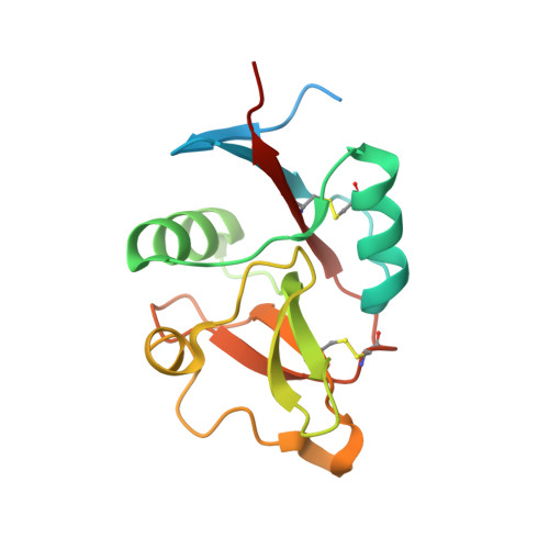
    4AK8

  • PubMed Abstract: 

    Langerin, a trimeric C-type lectin specifically expressed in Langerhans cells, has been reported to be a pathogen receptor through the recognition of glycan motifs by its three carbohydrate recognition domains (CRD). In the context of HIV-1 (human immunodeficiency virus-1) transmission, Langerhans cells of genital mucosa play a protective role by internalizing virions in Birbeck Granules (BG) for elimination. Langerin (Lg) is directly involved in virion binding and BG formation through its CRDs. However, nothing is known regarding the mechanism of langerin assembly underlying BG formation. We investigated at the molecular level the impact of two CRD mutations, W264R and F241L, on langerin structure, function, and BG assembly using a combination of biochemical and biophysical approaches. Although the W264R mutation causes CRD global unfolding, the F241L mutation does not affect the overall structure and gp120 (surface HIV-1 glycoprotein of 120 kDa) binding capacities of isolated Lg-CRD. In contrast, this mutation induces major functional and structural alterations of the whole trimeric langerin extracellular domain (Lg-ECD). As demonstrated by small-angle x-ray scattering comparative analysis of wild-type and mutant forms, the F241L mutation perturbs the oligomerization state and the global architecture of Lg-ECD. Correlatively, despite conserved intrinsic lectin activity of the CRD, avidity property of Lg-ECD is affected as shown by a marked decrease of gp120 binding. Beyond the change of residue itself, the F241L mutation induces relocation of the K200 side chain also located within the interface between protomers of trimeric Lg-ECD, thereby explaining the defective oligomerization of mutant Lg. We conclude that not only functional CRDs but also their correct spatial presentation are critical for BG formation as well as gp120 binding.


  • Organizational Affiliation
    • University Grenoble Alpes, IBS, Grenoble, France; CNRS, UMR 5075, Grenoble France; CEA, UMR 5075, Grenoble France.

Macromolecule Content 

  • Total Structure Weight: 71.85 kDa 
  • Atom Count: 5,148 
  • Modeled Residue Count: 521 
  • Deposited Residue Count: 620 
  • Unique protein chains: 1

Macromolecules

Find similar proteins by:|  3D Structure
Entity ID: 1
MoleculeChains  Sequence LengthOrganismDetailsImage
C-TYPE LECTIN DOMAIN FAMILY 4 MEMBER K
A, B, C, D
155Homo sapiensMutation(s): 2 
UniProt & NIH Common Fund Data Resources
Find proteins for Q9UJ71 (Homo sapiens)
Explore Q9UJ71 
Go to UniProtKB:  Q9UJ71
PHAROS:  Q9UJ71
GTEx:  ENSG00000116031 
Entity Groups
Sequence Clusters30% Identity50% Identity70% Identity90% Identity95% Identity100% Identity
UniProt GroupQ9UJ71
Sequence Annotations
Expand
Reference Sequence

Small Molecules

Ligands 3 Unique
IDChains Name / Formula / InChI Key2D Diagram3D Interactions
CA

Query on CA


Download:Ideal Coordinates CCD File
E [auth A],
G [auth B],
I [auth C],
L [auth D]
CALCIUM ION
Ca
BHPQYMZQTOCNFJ-UHFFFAOYSA-N
CL

Query on CL


Download:Ideal Coordinates CCD File
K [auth D]CHLORIDE ION
Cl
VEXZGXHMUGYJMC-UHFFFAOYSA-M
MG

Query on MG


Download:Ideal Coordinates CCD File
F [auth A],
H [auth B],
J [auth C],
M [auth D]
MAGNESIUM ION
Mg
JLVVSXFLKOJNIY-UHFFFAOYSA-N

Experimental Data & Validation

Experimental Data

  • Method: X-RAY DIFFRACTION
  • Resolution: 1.40 Å
  • R-Value Free:  0.219 (Depositor), 0.169 (DCC) 
  • R-Value Work:  0.175 (Depositor), 0.177 (DCC) 
  • R-Value Observed: 0.178 (Depositor) 
Space Group: P 42
Unit Cell:
Length ( Å )Angle ( ˚ )
a = 79.959α = 90
b = 79.959β = 90
c = 90.419γ = 90
Software Package:
Software NamePurpose
REFMACrefinement
XDSdata reduction
SCALAdata scaling
MOLREPphasing

Structure Validation

View Full Validation Report


Entry History 

Revision History  (Full details and data files)

  • Version 1.0: 2013-04-03
    Type: Initial release
  • Version 1.1: 2015-02-11
    Changes: Database references
  • Version 1.2: 2015-02-25
    Changes: Database references
  • Version 1.3: 2019-05-08
    Changes: Data collection, Experimental preparation, Other
  • Version 1.4: 2023-12-20
    Changes: Data collection, Database references, Derived calculations, Other, Refinement description
  • Version 1.5: 2024-10-23
    Changes: Structure summary