3WXF | pdb_00003wxf

Crystal structure of CYLD USP domain (C596S E674Q) in complex with Met1-linked diubiquitin


Experimental Data Snapshot

  • Method: X-RAY DIFFRACTION
  • Resolution: 2.30 Å
  • R-Value Free: 
    0.229 (Depositor), 0.228 (DCC) 
  • R-Value Work: 
    0.187 (Depositor), 0.191 (DCC) 
  • R-Value Observed: 
    0.189 (Depositor) 

Starting Models: experimental
View more details

wwPDB Validation 3D Report Full Report

Validation slider image for 3WXF

This is version 1.3 of the entry. See complete history

Literature

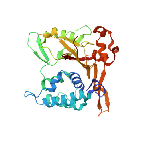
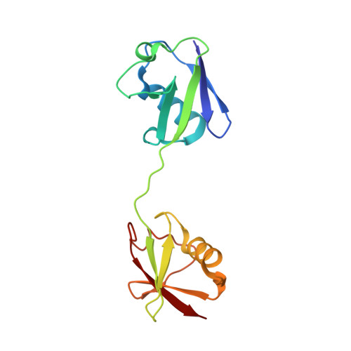
Structures of CYLD USP with Met1- or Lys63-linked diubiquitin reveal mechanisms for dual specificity.

Sato, Y.Goto, E.Shibata, Y.Kubota, Y.Yamagata, A.Goto-Ito, S.Kubota, K.Inoue, J.Takekawa, M.Tokunaga, F.Fukai, S.

(2015) Nat Struct Mol Biol 22: 222-229

  • DOI: https://doi.org/10.1038/nsmb.2970
  • Primary Citation Related Structures: 
    3WXE, 3WXF, 3WXG

  • PubMed Abstract: 

    The tumor suppressor CYLD belongs to a ubiquitin (Ub)-specific protease (USP) family and specifically cleaves Met1- and Lys63-linked polyubiquitin chains to suppress inflammatory signaling pathways. Here, we report crystal structures representing the catalytic states of zebrafish CYLD for Met1- and Lys63-linked Ub chains and two distinct precatalytic states for Met1-linked chains. In both catalytic states, the distal Ub is bound to CYLD in a similar manner, and the scissile bond is located close to the catalytic residue, whereas the proximal Ub is bound in a manner specific to Met1- or Lys63-linked chains. Further structure-based mutagenesis experiments support the mechanism by which CYLD specifically cleaves both Met1- and Lys63-linked chains and provide insight into tumor-associated mutations of CYLD. This study provides new structural insight into the mechanisms by which USP family deubiquitinating enzymes recognize and cleave Ub chains with specific linkage types.


  • Organizational Affiliation
    • 1] Life Science Division, Synchrotron Radiation Research Organization, University of Tokyo, Tokyo, Japan. [2] Center for Structural Biology of Challenging Proteins, Institute of Molecular and Cellular Biosciences, University of Tokyo, Tokyo, Japan. [3] Department of Medical Genome Sciences, Graduate School of Frontier Sciences, University of Tokyo, Chiba, Japan.

Macromolecule Content 

  • Total Structure Weight: 105.68 kDa 
  • Atom Count: 7,358 
  • Modeled Residue Count: 894 
  • Deposited Residue Count: 920 
  • Unique protein chains: 2

Macromolecules

Find similar proteins by:|  3D Structure
Entity ID: 1
MoleculeChains  Sequence LengthOrganismDetailsImage
Uncharacterized protein
A, C
312Danio rerioMutation(s): 2 
Gene Names: cyldcylda
EC: 3.4.19.12
UniProt
Find proteins for E7FEV5 (Danio rerio)
Explore E7FEV5 
Go to UniProtKB:  E7FEV5
Entity Groups
Sequence Clusters30% Identity50% Identity70% Identity90% Identity95% Identity100% Identity
UniProt GroupE7FEV5
Sequence Annotations
Expand
Reference Sequence
Find similar proteins by:|  3D Structure
Entity ID: 2
MoleculeChains  Sequence LengthOrganismDetailsImage
Ubiquitin
B, D
148Homo sapiensMutation(s): 0 
Gene Names: UBC
UniProt & NIH Common Fund Data Resources
Find proteins for P0CG48 (Homo sapiens)
Explore P0CG48 
Go to UniProtKB:  P0CG48
PHAROS:  P0CG48
GTEx:  ENSG00000150991 
Entity Groups
Sequence Clusters30% Identity50% Identity70% Identity90% Identity95% Identity100% Identity
UniProt GroupP0CG48
Sequence Annotations
Expand
Reference Sequence

Experimental Data & Validation

Experimental Data

  • Method: X-RAY DIFFRACTION
  • Resolution: 2.30 Å
  • R-Value Free:  0.229 (Depositor), 0.228 (DCC) 
  • R-Value Work:  0.187 (Depositor), 0.191 (DCC) 
  • R-Value Observed: 0.189 (Depositor) 
Space Group: P 1
Unit Cell:
Length ( Å )Angle ( ˚ )
a = 49.628α = 77.66
b = 65.372β = 89.04
c = 69.846γ = 89.46
Software Package:
Software NamePurpose
MOLREPphasing
REFMACrefinement
HKL-2000data reduction
HKL-2000data scaling

Structure Validation

View Full Validation Report


Entry History 

Deposition Data

Revision History  (Full details and data files)

  • Version 1.0: 2015-02-11
    Type: Initial release
  • Version 1.1: 2017-08-16
    Changes: Refinement description, Source and taxonomy
  • Version 1.2: 2020-01-01
    Changes: Database references
  • Version 1.3: 2023-11-08
    Changes: Data collection, Database references, Derived calculations, Refinement description