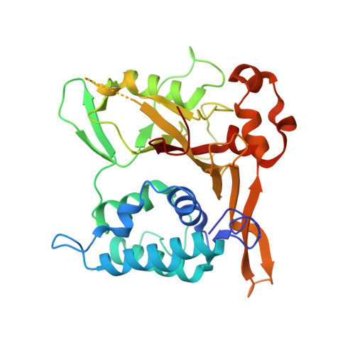
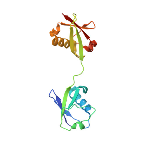
3WXE

Crystal structure of CYLD USP domain (C596S) in complex with Met1-linked diubiquitin


Experimental Data Snapshot

  • Method: X-RAY DIFFRACTION
  • Resolution: 2.50 Å
  • R-Value Free: 0.247 
  • R-Value Work: 0.182 
  • R-Value Observed: 0.185 

wwPDB Validation   3D Report Full Report


This is version 1.3 of the entry. See complete history


Literature

Structures of CYLD USP with Met1- or Lys63-linked diubiquitin reveal mechanisms for dual specificity.

Sato, Y.Goto, E.Shibata, Y.Kubota, Y.Yamagata, A.Goto-Ito, S.Kubota, K.Inoue, J.Takekawa, M.Tokunaga, F.Fukai, S.

(2015) Nat Struct Mol Biol 22: 222-229

  • DOI: https://doi.org/10.1038/nsmb.2970
  • Primary Citation of Related Structures:  
    3WXE, 3WXF, 3WXG

  • PubMed Abstract: 

    The tumor suppressor CYLD belongs to a ubiquitin (Ub)-specific protease (USP) family and specifically cleaves Met1- and Lys63-linked polyubiquitin chains to suppress inflammatory signaling pathways. Here, we report crystal structures representing the catalytic states of zebrafish CYLD for Met1- and Lys63-linked Ub chains and two distinct precatalytic states for Met1-linked chains. In both catalytic states, the distal Ub is bound to CYLD in a similar manner, and the scissile bond is located close to the catalytic residue, whereas the proximal Ub is bound in a manner specific to Met1- or Lys63-linked chains. Further structure-based mutagenesis experiments support the mechanism by which CYLD specifically cleaves both Met1- and Lys63-linked chains and provide insight into tumor-associated mutations of CYLD. This study provides new structural insight into the mechanisms by which USP family deubiquitinating enzymes recognize and cleave Ub chains with specific linkage types.


  • Organizational Affiliation

    1] Life Science Division, Synchrotron Radiation Research Organization, University of Tokyo, Tokyo, Japan. [2] Center for Structural Biology of Challenging Proteins, Institute of Molecular and Cellular Biosciences, University of Tokyo, Tokyo, Japan. [3] Department of Medical Genome Sciences, Graduate School of Frontier Sciences, University of Tokyo, Chiba, Japan.


Macromolecules
Find similar proteins by:  (by identity cutoff)  |  3D Structure
Entity ID: 1
MoleculeChains Sequence LengthOrganismDetailsImage
Uncharacterized protein312Danio rerioMutation(s): 1 
Gene Names: cyldcylda
UniProt
Find proteins for E7FEV5 (Danio rerio)
Explore E7FEV5 
Go to UniProtKB:  E7FEV5
Entity Groups  
Sequence Clusters30% Identity50% Identity70% Identity90% Identity95% Identity100% Identity
UniProt GroupE7FEV5
Sequence Annotations
Expand
  • Reference Sequence
Find similar proteins by:  (by identity cutoff)  |  3D Structure
Entity ID: 2
MoleculeChains Sequence LengthOrganismDetailsImage
Ubiquitin148Homo sapiensMutation(s): 0 
Gene Names: UBC
UniProt & NIH Common Fund Data Resources
Find proteins for P0CG48 (Homo sapiens)
Explore P0CG48 
Go to UniProtKB:  P0CG48
PHAROS:  P0CG48
GTEx:  ENSG00000150991 
Entity Groups  
Sequence Clusters30% Identity50% Identity70% Identity90% Identity95% Identity100% Identity
UniProt GroupP0CG48
Sequence Annotations
Expand
  • Reference Sequence
Experimental Data & Validation

Experimental Data

  • Method: X-RAY DIFFRACTION
  • Resolution: 2.50 Å
  • R-Value Free: 0.247 
  • R-Value Work: 0.182 
  • R-Value Observed: 0.185 
  • Space Group: P 1 21 1
Unit Cell:
Length ( Å )Angle ( ˚ )
a = 64.676α = 90
b = 49.788β = 102.69
c = 71.003γ = 90
Software Package:
Software NamePurpose
MOLREPphasing
REFMACrefinement
HKL-2000data reduction
HKL-2000data scaling

Structure Validation

View Full Validation Report


Entry History 

Deposition Data

Revision History  (Full details and data files)

  • Version 1.0: 2015-02-11
    Type: Initial release
  • Version 1.1: 2017-08-16
    Changes: Refinement description, Source and taxonomy
  • Version 1.2: 2020-01-01
    Changes: Database references
  • Version 1.3: 2023-11-08
    Changes: Data collection, Database references, Refinement description