3W82

Human alpha-L-iduronidase in complex with iduronic acid


Experimental Data Snapshot

  • Method: X-RAY DIFFRACTION
  • Resolution: 2.76 Å
  • R-Value Free: 0.254 
  • R-Value Work: 0.195 
  • R-Value Observed: 0.198 

wwPDB Validation   3D Report Full Report


Ligand Structure Quality Assessment 


This is version 2.0 of the entry. See complete history


Literature

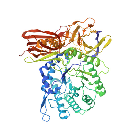
Human alpha-L-iduronidase uses its own N-glycan as a substrate-binding and catalytic module

Maita, N.Tsukimura, T.Taniguchi, T.Saito, S.Ohno, K.Taniguchi, H.Sakuraba, H.

(2013) Proc Natl Acad Sci U S A 110: 14628-14633

  • DOI: https://doi.org/10.1073/pnas.1306939110
  • Primary Citation of Related Structures:  
    3W81, 3W82

  • PubMed Abstract: 
  • N-glycosylation is a major posttranslational modification that endows proteins with various functions. It is established that N-glycans are essential for the correct folding and stability of some enzymes; however, the actual effects of N-glycans on their activities are poorly understood ...

    N-glycosylation is a major posttranslational modification that endows proteins with various functions. It is established that N-glycans are essential for the correct folding and stability of some enzymes; however, the actual effects of N-glycans on their activities are poorly understood. Here, we show that human α-l-iduronidase (hIDUA), of which a dysfunction causes accumulation of dermatan/heparan sulfate leading to mucopolysaccharidosis type I, uses its own N-glycan as a substrate binding and catalytic module. Structural analysis revealed that the mannose residue of the N-glycan attached to N372 constituted a part of the substrate-binding pocket and interacted directly with a substrate. A deglycosylation study showed that enzyme activity was highly correlated with the N-glycan attached to N372. The kinetics of native and deglycosylated hIDUA suggested that the N-glycan is also involved in catalytic processes. Our study demonstrates a previously unrecognized function of N-glycans.


    Organizational Affiliation

    Laboratory of X-Ray Crystallography, Institute for Enzyme Research, University of Tokushima, Tokushima 770-8503, Japan. nmaita@tokushima-u.ac.jp


Macromolecules
Find similar proteins by:  (by identity cutoff)  |  3D Structure
Entity ID: 1
MoleculeChainsSequence LengthOrganismDetailsImage
Alpha-L-iduronidaseA, B627Homo sapiensMutation(s): 0 
Gene Names: IDUA
EC: 3.2.1.76
UniProt & NIH Common Fund Data Resources
Find proteins for P35475 (Homo sapiens)
Explore P35475 
Go to UniProtKB:  P35475
PHAROS:  P35475
Entity Groups  
Sequence Clusters30% Identity50% Identity70% Identity90% Identity95% Identity100% Identity
UniProt GroupP35475
Protein Feature View
Expand
  • Reference Sequence
Oligosaccharides

Help

Entity ID: 2
MoleculeChainsChain Length2D DiagramGlycosylation3D Interactions
alpha-D-mannopyranose-(1-2)-alpha-D-mannopyranose-(1-3)-[alpha-D-mannopyranose-(1-2)-alpha-D-mannopyranose-(1-6)]alpha-D-mannopyranose-(1-6)-[alpha-D-mannopyranose-(1-2)-alpha-D-mannopyranose-(1-3)]beta-D-mannopyranose-(1-4)-2-acetamido-2-deoxy-beta-D-glucopyranose-(1-4)-2-acetamido-2-deoxy-beta-D-glucopyranoseC 10N-Glycosylation Oligosaccharides Interaction
Glycosylation Resources
GlyTouCan:  G89864BN
GlyCosmos:  G89864BN
GlyGen:  G89864BN
Entity ID: 3
MoleculeChainsChain Length2D DiagramGlycosylation3D Interactions
2-acetamido-2-deoxy-beta-D-glucopyranose-(1-4)-2-acetamido-2-deoxy-beta-D-glucopyranoseD, G 2N-Glycosylation Oligosaccharides Interaction
Glycosylation Resources
GlyTouCan:  G42666HT
GlyCosmos:  G42666HT
GlyGen:  G42666HT
Entity ID: 4
MoleculeChainsChain Length2D DiagramGlycosylation3D Interactions
alpha-D-mannopyranose-(1-2)-alpha-D-mannopyranose-(1-6)-[alpha-D-mannopyranose-(1-3)]alpha-D-mannopyranose-(1-6)-[alpha-D-mannopyranose-(1-2)-alpha-D-mannopyranose-(1-3)]beta-D-mannopyranose-(1-4)-2-acetamido-2-deoxy-beta-D-glucopyranose-(1-4)-2-acetamido-2-deoxy-beta-D-glucopyranoseE 9N-Glycosylation Oligosaccharides Interaction
Glycosylation Resources
GlyTouCan:  G83161QT
GlyCosmos:  G83161QT
GlyGen:  G83161QT
Entity ID: 5
MoleculeChainsChain Length2D DiagramGlycosylation3D Interactions
alpha-L-fucopyranose-(1-6)-2-acetamido-2-deoxy-beta-D-glucopyranoseF 2N-Glycosylation Oligosaccharides Interaction
Glycosylation Resources
GlyTouCan:  G86851RC
GlyCosmos:  G86851RC
GlyGen:  G86851RC
Small Molecules
Ligands 6 Unique
IDChainsName / Formula / InChI Key2D Diagram3D Interactions
NAG
Query on NAG

Download Ideal Coordinates CCD File 
H [auth A],
L [auth B]
2-acetamido-2-deoxy-beta-D-glucopyranose
C8 H15 N O6
OVRNDRQMDRJTHS-FMDGEEDCSA-N
 Ligand Interaction
IDR
Query on IDR

Download Ideal Coordinates CCD File 
I [auth A],
M [auth B]
alpha-L-idopyranuronic acid
C6 H10 O7
AEMOLEFTQBMNLQ-VCSGLWQLSA-N
 Ligand Interaction
TLA
Query on TLA

Download Ideal Coordinates CCD File 
N [auth B]L(+)-TARTARIC ACID
C4 H6 O6
FEWJPZIEWOKRBE-JCYAYHJZSA-N
 Ligand Interaction
PO4
Query on PO4

Download Ideal Coordinates CCD File 
J [auth A],
P [auth B]
PHOSPHATE ION
O4 P
NBIIXXVUZAFLBC-UHFFFAOYSA-K
 Ligand Interaction
PO3
Query on PO3

Download Ideal Coordinates CCD File 
O [auth B]PHOSPHITE ION
O3 P
AQSJGOWTSHOLKH-UHFFFAOYSA-N
 Ligand Interaction
CL
Query on CL

Download Ideal Coordinates CCD File 
K [auth A],
Q [auth B]
CHLORIDE ION
Cl
VEXZGXHMUGYJMC-UHFFFAOYSA-M
 Ligand Interaction
Experimental Data & Validation

Experimental Data

  • Method: X-RAY DIFFRACTION
  • Resolution: 2.76 Å
  • R-Value Free: 0.254 
  • R-Value Work: 0.195 
  • R-Value Observed: 0.198 
  • Space Group: H 3
Unit Cell:
Length ( Å )Angle ( ˚ )
a = 259.98α = 90
b = 259.98β = 90
c = 65.3γ = 120
Software Package:
Software NamePurpose
ADSCdata collection
MOLREPphasing
REFMACrefinement
MOSFLMdata reduction
Aimlessdata scaling

Structure Validation

View Full Validation Report


Ligand Structure Quality Assessment 


Entry History 

Deposition Data

Revision History  (Full details and data files)

  • Version 1.0: 2013-08-21
    Type: Initial release
  • Version 1.1: 2013-10-16
    Changes: Database references
  • Version 2.0: 2020-07-29
    Type: Remediation
    Reason: Carbohydrate remediation
    Changes: Atomic model, Data collection, Derived calculations, Structure summary