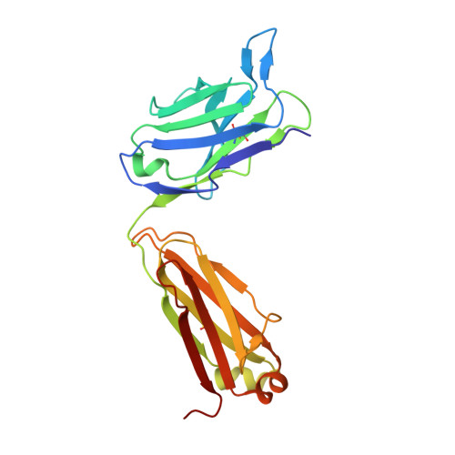
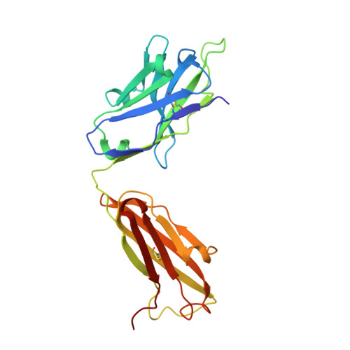
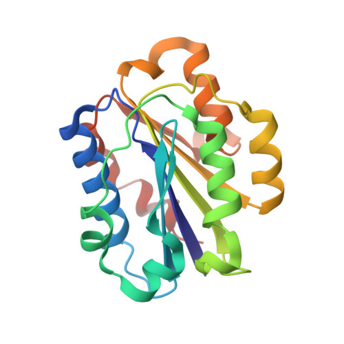
3QA3

Crystal Structure of A-domain in complex with antibody


Experimental Data Snapshot

  • Method: X-RAY DIFFRACTION
  • Resolution: 3.00 Å
  • R-Value Free: 0.224 
  • R-Value Work: 0.194 
  • R-Value Observed: 0.196 

Starting Model: experimental
View more details

wwPDB Validation   3D Report Full Report


This is version 1.3 of the entry. See complete history


Literature

Stable Coordination of the Inhibitory Ca2+ Ion at the Metal Ion-Dependent Adhesion Site in Integrin CD11b/CD18 by an Antibody-Derived Ligand Aspartate: Implications for Integrin Regulation and Structure-Based Drug Design.

Mahalingam, B.Ajroud, K.Alonso, J.L.Anand, S.Adair, B.D.Horenstein, A.L.Malavasi, F.Xiong, J.P.Arnaout, M.A.

(2011) J Immunol 187: 6393-6401

  • DOI: https://doi.org/10.4049/jimmunol.1102394
  • Primary Citation of Related Structures:  
    3Q3G, 3QA3

  • PubMed Abstract: 

    A central feature of integrin interaction with physiologic ligands is the monodentate binding of a ligand carboxylate to a Mg(2+) ion hexacoordinated at the metal ion-dependent adhesion site (MIDAS) in the integrin A domain. This interaction stabilizes the A domain in the high-affinity state, which is distinguished from the default low-affinity state by tertiary changes in the domain that culminate in cell adhesion. Small molecule ligand-mimetic integrin antagonists act as partial agonists, eliciting similar activating conformational changes in the A domain, which has contributed to paradoxical adhesion and increased patient mortality in large clinical trials. As with other ligand-mimetic integrin antagonists, the function-blocking mAb 107 binds MIDAS of integrin CD11b/CD18 A domain (CD11bA), but in contrast, it favors the inhibitory Ca(2+) ion over the Mg(2+) ion at MIDAS. We determined the crystal structures of the Fab fragment of mAb 107 complexed to the low- and high-affinity states of CD11bA. Favored binding of the Ca(2+) ion at MIDAS is caused by the unusual symmetric bidentate ligation of a Fab-derived ligand Asp to a heptacoordinated MIDAS Ca(2+) ion. Binding of the Fab fragment of mAb 107 to CD11bA did not trigger the activating tertiary changes in the domain or in the full-length integrin. These data show that the denticity of the ligand Asp/Glu can modify the divalent cation selectivity at MIDAS and hence integrin function. Stabilizing the Ca(2+) ion at MIDAS by bidentate ligation to a ligand Asp/Glu may provide one approach for designing pure integrin antagonists.


  • Organizational Affiliation

    Structural Biology Program, Division of Nephrology, Massachusetts General Hospital, Harvard Medical School, Charlestown, MA 02129, USA.


Macromolecules
Find similar proteins by:  (by identity cutoff)  |  3D Structure
Entity ID: 1
MoleculeChains Sequence LengthOrganismDetailsImage
Antibody Light chainA [auth C],
D [auth A],
G [auth F],
J
220Mus musculusMutation(s): 0 
Entity Groups  
Sequence Clusters30% Identity50% Identity70% Identity90% Identity95% Identity100% Identity
Sequence Annotations
Expand
  • Reference Sequence
Find similar proteins by:  (by identity cutoff)  |  3D Structure
Entity ID: 2
MoleculeChains Sequence LengthOrganismDetailsImage
Antibody Heavy chainB [auth D],
E [auth B],
H,
K
224Mus musculusMutation(s): 0 
Entity Groups  
Sequence Clusters30% Identity50% Identity70% Identity90% Identity95% Identity100% Identity
Sequence Annotations
Expand
  • Reference Sequence
Find similar proteins by:  (by identity cutoff)  |  3D Structure
Entity ID: 3
MoleculeChains Sequence LengthOrganismDetailsImage
Integrin alpha-MC [auth G],
F [auth E],
I,
L
190Homo sapiensMutation(s): 1 
Gene Names: ITGAMCD11BCR3A
UniProt & NIH Common Fund Data Resources
Find proteins for P11215 (Homo sapiens)
Explore P11215 
Go to UniProtKB:  P11215
PHAROS:  P11215
GTEx:  ENSG00000169896 
Entity Groups  
Sequence Clusters30% Identity50% Identity70% Identity90% Identity95% Identity100% Identity
UniProt GroupP11215
Sequence Annotations
Expand
  • Reference Sequence
Small Molecules
Ligands 3 Unique
IDChains Name / Formula / InChI Key2D Diagram3D Interactions
GOL
Query on GOL

Download Ideal Coordinates CCD File 
O [auth D],
S [auth G],
T [auth G],
U [auth A],
V [auth B]
GLYCEROL
C3 H8 O3
PEDCQBHIVMGVHV-UHFFFAOYSA-N
EDO
Query on EDO

Download Ideal Coordinates CCD File 
AA [auth J]
BA [auth K]
M [auth C]
N [auth C]
P [auth D]
AA [auth J],
BA [auth K],
M [auth C],
N [auth C],
P [auth D],
Q [auth D],
X [auth H],
Z [auth J]
1,2-ETHANEDIOL
C2 H6 O2
LYCAIKOWRPUZTN-UHFFFAOYSA-N
CA
Query on CA

Download Ideal Coordinates CCD File 
CA [auth L],
R [auth G],
W [auth E],
Y [auth I]
CALCIUM ION
Ca
BHPQYMZQTOCNFJ-UHFFFAOYSA-N
Experimental Data & Validation

Experimental Data

  • Method: X-RAY DIFFRACTION
  • Resolution: 3.00 Å
  • R-Value Free: 0.224 
  • R-Value Work: 0.194 
  • R-Value Observed: 0.196 
  • Space Group: P 21 21 21
Unit Cell:
Length ( Å )Angle ( ˚ )
a = 82.604α = 90
b = 158.346β = 90
c = 233.319γ = 90
Software Package:
Software NamePurpose
BUSTERrefinement
HKL-2000data reduction
HKL-2000data scaling
PHASERphasing

Structure Validation

View Full Validation Report


Entry History 

Deposition Data

Revision History  (Full details and data files)

  • Version 1.0: 2011-11-30
    Type: Initial release
  • Version 1.1: 2011-12-28
    Changes: Database references
  • Version 1.2: 2023-09-13
    Changes: Data collection, Database references, Derived calculations, Refinement description
  • Version 1.3: 2024-10-09
    Changes: Structure summary