3BEF

Crystal structure of thrombin bound to the extracellular fragment of PAR1


Experimental Data Snapshot

  • Method: X-RAY DIFFRACTION
  • Resolution: 2.20 Å
  • R-Value Free: 0.248 
  • R-Value Work: 0.207 
  • R-Value Observed: 0.207 

wwPDB Validation   3D Report Full Report


This is version 1.4 of the entry. See complete history


Literature

Structural identification of the pathway of long-range communication in an allosteric enzyme.

Gandhi, P.S.Chen, Z.Mathews, F.S.Di Cera, E.

(2008) Proc Natl Acad Sci U S A 105: 1832-1837

  • DOI: https://doi.org/10.1073/pnas.0710894105
  • Primary Citation of Related Structures:  
    3BEF, 3BEI

  • PubMed Abstract: 

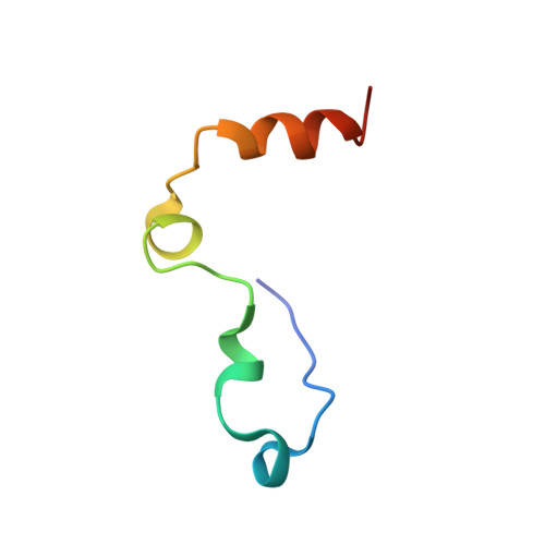
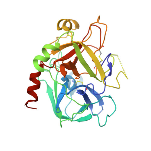
    Allostery is a common mechanism of regulation of enzyme activity and specificity, and its signatures are readily identified from functional studies. For many allosteric systems, structural evidence exists of long-range communication among protein domains, but rarely has this communication been traced to a detailed pathway. The thrombin mutant D102N is stabilized in a self-inhibited conformation where access to the active site is occluded by a collapse of the entire 215-219 beta-strand. Binding of a fragment of the protease activated receptor PAR1 to exosite I, 30-A away from the active site region, causes a large conformational change that corrects the position of the 215-219 beta-strand and restores access to the active site. The crystal structure of the thrombin-PAR1 complex, solved at 2.2-A resolution, reveals the details of this long-range allosteric communication in terms of a network of polar interactions.


  • Organizational Affiliation

    Department of Biochemistry and Molecular Biophysics, Washington University School of Medicine, Box 8231, St. Louis, MO 63110, USA.


Macromolecules
Find similar proteins by:  (by identity cutoff)  |  3D Structure
Entity ID: 1
MoleculeChains Sequence LengthOrganismDetailsImage
Prothrombin
A, D
46Homo sapiensMutation(s): 0 
Gene Names: F2
EC: 3.4.21.5
UniProt & NIH Common Fund Data Resources
Find proteins for P00734 (Homo sapiens)
Explore P00734 
Go to UniProtKB:  P00734
PHAROS:  P00734
GTEx:  ENSG00000180210 
Entity Groups  
Sequence Clusters30% Identity50% Identity70% Identity90% Identity95% Identity100% Identity
UniProt GroupP00734
Sequence Annotations
Expand
  • Reference Sequence
Find similar proteins by:  (by identity cutoff)  |  3D Structure
Entity ID: 2
MoleculeChains Sequence LengthOrganismDetailsImage
Prothrombin
B, E
259Homo sapiensMutation(s): 1 
Gene Names: F2
EC: 3.4.21.5
UniProt & NIH Common Fund Data Resources
Find proteins for P00734 (Homo sapiens)
Explore P00734 
Go to UniProtKB:  P00734
PHAROS:  P00734
GTEx:  ENSG00000180210 
Entity Groups  
Sequence Clusters30% Identity50% Identity70% Identity90% Identity95% Identity100% Identity
UniProt GroupP00734
Sequence Annotations
Expand
  • Reference Sequence

Find similar proteins by:  Sequence   |   3D Structure  

Entity ID: 3
MoleculeChains Sequence LengthOrganismDetailsImage
Proteinase-activated receptor 1
C, F
9Homo sapiensMutation(s): 0 
Gene Names: F2RCF2RPAR1TR
UniProt & NIH Common Fund Data Resources
Find proteins for P25116 (Homo sapiens)
Explore P25116 
Go to UniProtKB:  P25116
PHAROS:  P25116
GTEx:  ENSG00000181104 
Entity Groups  
UniProt GroupP25116
Sequence Annotations
Expand
  • Reference Sequence
Small Molecules
Ligands 1 Unique
IDChains Name / Formula / InChI Key2D Diagram3D Interactions
NAG
Query on NAG

Download Ideal Coordinates CCD File 
G [auth B],
H [auth E]
2-acetamido-2-deoxy-beta-D-glucopyranose
C8 H15 N O6
OVRNDRQMDRJTHS-FMDGEEDCSA-N
Experimental Data & Validation

Experimental Data

  • Method: X-RAY DIFFRACTION
  • Resolution: 2.20 Å
  • R-Value Free: 0.248 
  • R-Value Work: 0.207 
  • R-Value Observed: 0.207 
  • Space Group: P 1
Unit Cell:
Length ( Å )Angle ( ˚ )
a = 46.06α = 76.89
b = 50.342β = 84.3
c = 85.078γ = 73.69
Software Package:
Software NamePurpose
CNSrefinement
HKL-2000data collection
HKL-2000data reduction
SCALEPACKdata scaling
MOLREPphasing

Structure Validation

View Full Validation Report


Entry History 

Deposition Data

Revision History  (Full details and data files)

  • Version 1.0: 2008-01-01
    Type: Initial release
  • Version 1.1: 2011-07-13
    Changes: Non-polymer description, Version format compliance
  • Version 1.2: 2020-07-29
    Type: Remediation
    Reason: Carbohydrate remediation
    Changes: Data collection, Database references, Derived calculations, Structure summary
  • Version 1.3: 2021-10-20
    Changes: Database references, Structure summary
  • Version 1.4: 2023-08-30
    Changes: Data collection, Refinement description