1ZEB | pdb_00001zeb

X-ray structure of alkaline phosphatase from human placenta in complex with 5'-AMP


Experimental Data Snapshot

  • Method: X-RAY DIFFRACTION
  • Resolution: 1.90 Å
  • R-Value Free: 
    0.197 (Depositor) 
  • R-Value Work: 
    0.151 (Depositor) 
  • R-Value Observed: 
    0.153 (Depositor) 

wwPDB Validation 3D Report Full Report

Validation slider image for 1ZEB

Ligand Structure Quality Assessment 


This is version 1.4 of the entry. See complete history

Re-refinement Note

A newer entry is available that reflects an alternative modeling of the original data: 3MK0

Literature

Structural Studies of Human Placental Alkaline Phosphatase in Complex with Functional Ligands.

Llinas, P.Stura, E.A.Menez, A.Kiss, Z.Stigbrand, T.Millan, J.L.Le Du, M.H.

(2005) J Mol Biology 350: 441-451

  • DOI: https://doi.org/10.1016/j.jmb.2005.04.068
  • Primary Citation Related Structures: 
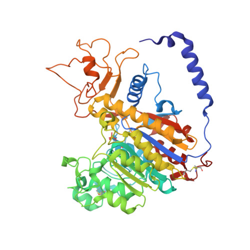
    1ZEB, 1ZED, 1ZEF

  • PubMed Abstract: 

    The activity of human placental alkaline phosphatase (PLAP) is downregulated by a number of effectors such as l-phenylalanine, an uncompetitive inhibitor, 5'-AMP, an antagonist of the effects of PLAP on fibroblast proliferation and by p-nitrophenyl-phosphonate (PNPPate), a non-hydrolysable substrate analogue. For the first two, such regulation may be linked to its biological function that requires a reduced and better-regulated hydrolytic rate. To understand how such disparate ligands are able to inhibit the enzyme, we solved the structure of the complexes at 1.6A, 1.9A and 1.9A resolution, respectively. These crystal structures are the first of an alkaline phosphatase in complex with organic inhibitors. Of the three inhibitors, only l-Phe and PNPPate bind at the active site hydrophobic pocket, providing structural data on the uncompetitive inhibition process. In contrast, all three ligands interact at a remote peripheral site located 28A from the active site. In order to extend these observations to the other members of the human alkaline phosphatase family, we have modelled the structures of the other human isozymes and compared them to PLAP. This comparison highlights the crucial role played by position 429 at the active site in the modulation of the catalytic process, and suggests that the peripheral binding site may be involved in the functional specialization of the PLAP isozyme.


  • Organizational Affiliation
    • Laboratoire de Structure des Protéines, Département d'Ingénierie et d'Etude des Protéines, Commissariat à l'Energie Atomique, 91191 Gif sur Yvette, France.

Macromolecule Content 

  • Total Structure Weight: 53.53 kDa 
  • Atom Count: 4,419 
  • Modeled Residue Count: 480 
  • Deposited Residue Count: 484 
  • Unique protein chains: 1

Macromolecules

Find similar proteins by:|  3D Structure
Entity ID: 1
MoleculeChains  Sequence LengthOrganismDetailsImage
Alkaline phosphatase484Homo sapiensMutation(s): 0 
EC: 3.1.3.1
UniProt & NIH Common Fund Data Resources
Find proteins for P05187 (Homo sapiens)
Explore P05187 
Go to UniProtKB:  P05187
PHAROS:  P05187
GTEx:  ENSG00000163283 
Entity Groups
Sequence Clusters30% Identity50% Identity70% Identity90% Identity95% Identity100% Identity
UniProt GroupP05187
Glycosylation
Glycosylation Sites: 2Go to GlyGen: P05187-1
Sequence Annotations
Expand
Reference Sequence

Small Molecules

Ligands 4 Unique
IDChains Name / Formula / InChI Key2D Diagram3D Interactions
NAG

Query on NAG


Download:Ideal Coordinates CCD File
B [auth A],
C [auth A]
2-acetamido-2-deoxy-beta-D-glucopyranose
C8 H15 N O6
OVRNDRQMDRJTHS-FMDGEEDCSA-N
ZN

Query on ZN


Download:Ideal Coordinates CCD File
D [auth A],
E [auth A]
ZINC ION
Zn
PTFCDOFLOPIGGS-UHFFFAOYSA-N
CA

Query on CA


Download:Ideal Coordinates CCD File
G [auth A]CALCIUM ION
Ca
BHPQYMZQTOCNFJ-UHFFFAOYSA-N
MG

Query on MG


Download:Ideal Coordinates CCD File
F [auth A]MAGNESIUM ION
Mg
JLVVSXFLKOJNIY-UHFFFAOYSA-N
Modified Residues  1 Unique
IDChains TypeFormula2D DiagramParent
SEP
Query on SEP
A
L-PEPTIDE LINKINGC3 H8 N O6 PSER

Experimental Data & Validation

Experimental Data

  • Method: X-RAY DIFFRACTION
  • Resolution: 1.90 Å
  • R-Value Free:  0.197 (Depositor) 
  • R-Value Work:  0.151 (Depositor) 
  • R-Value Observed: 0.153 (Depositor) 
Space Group: C 2 2 21
Unit Cell:
Length ( Å )Angle ( ˚ )
a = 89.022α = 90
b = 113.894β = 90
c = 106.594γ = 90
Software Package:
Software NamePurpose
REFMACrefinement
DENZOdata reduction
SCALEPACKdata scaling
FFTphasing

Structure Validation

View Full Validation Report


Ligand Structure Quality Assessment 


Entry History 

Deposition Data

Revision History  (Full details and data files)

  • Version 1.0: 2005-06-28
    Type: Initial release
  • Version 1.1: 2008-04-30
    Changes: Version format compliance
  • Version 1.2: 2011-07-13
    Changes: Derived calculations, Version format compliance
  • Version 1.3: 2020-07-29
    Type: Remediation
    Reason: Carbohydrate remediation
    Changes: Data collection, Derived calculations, Structure summary
  • Version 1.4: 2024-10-16
    Changes: Data collection, Database references, Structure summary