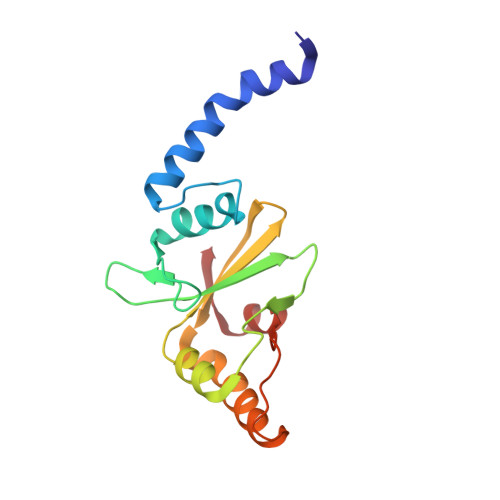
Structural and Biochemical Characterization of a New Mg(2+) Binding Site Near Tyr94 in the Restriction Endonuclease PvuII.
Spyridaki, A., Matzen, C., Lanio, T., Jeltsch, A., Simoncsits, A., Athanasiadis, A., Scheuring-Vanamee, E., Kokkinidis, M., Pingoud, A.(2003) J Mol Biology 331: 395
- PubMed: 12888347
- DOI: https://doi.org/10.1016/s0022-2836(03)00692-2
- Primary Citation of Related Structures:
1H56 - PubMed Abstract:
We have determined the crystal structure of the PvuII endonuclease in the presence of Mg(2+). According to the structural data, divalent metal ion binding in the PvuII subunits is highly asymmetric. The PvuII-Mg(2+) complex has two distinct metal ion binding sites, one in each monomer. One site is formed by the catalytic residues Asp58 and Glu68, and has extensive similarities to a catalytically important site found in all structurally examined restriction endonucleases. The other binding site is located in the other monomer, in the immediate vicinity of the hydroxyl group of Tyr94; it has no analogy to metal ion binding sites found so far in restriction endonucleases. To assign the number of metal ions involved and to better understand the role of Mg(2+) binding to Tyr94 for the function of PvuII, we have exchanged Tyr94 by Phe and characterized the metal ion dependence of DNA cleavage of wild-type PvuII and the Y94F variant. Wild-type PvuII cleaves both strands of the DNA in a concerted reaction. Mg(2+) binding, as measured by the Mg(2+) dependence of DNA cleavage, occurs with a Hill coefficient of 4, meaning that at least two metal ions are bound to each subunit in a cooperative fashion upon formation of the active complex. Quenched-flow experiments show that DNA cleavage occurs about tenfold faster if Mg(2+) is pre-incubated with enzyme or DNA than if preformed enzyme-DNA complexes are mixed with Mg(2+). These results show that Mg(2+) cannot easily enter the active center of the preformed enzyme-DNA complex, but that for fast cleavage the metal ions must already be bound to the apoenzyme and carried with the enzyme into the enzyme-DNA complex. The Y94F variant, in contrast to wild-type PvuII, does not cleave DNA in a concerted manner and metal ion binding occurs with a Hill coefficient of 1. These results indicate that removal of the Mg(2+) binding site at Tyr94 completely disrupts the cooperativity in DNA cleavage. Moreover, in quenched-flow experiments Y94F cleaves DNA about ten times more slowly than wild-type PvuII, regardless of the order of mixing. From these results we conclude that wild-type PvuII cleaves DNA in a fast and concerted reaction, because the Mg(2+) required for catalysis are already bound at the enzyme, one of them at Tyr94. We suggest that this Mg(2+) is shifted to the active center during binding of a specific DNA substrate. These results, for the first time, shed light on the pathway by which metal ions as essential cofactors enter the catalytic center of restriction endonucleases.
- Department of Biology, IMBB/FORTH, University of Crete, PO Box 1527, Heraklion, GR-71110, Crete, Greece.
Organizational Affiliation:


