1WAY | pdb_00001way

Active site thrombin inhibitors


Experimental Data Snapshot

  • Method: X-RAY DIFFRACTION
  • Resolution: 2.02 Å
  • R-Value Free: 
    0.231 (Depositor) 
  • R-Value Work: 
    0.158 (Depositor) 
  • R-Value Observed: 
    0.161 (Depositor) 

Starting Model: experimental
View more details

wwPDB Validation 3D Report Full Report

Validation slider image for 1WAY

This is version 1.4 of the entry. See complete history

Literature

Fragment-Based Lead Discovery Using X-Ray Crystallography

Hartshorn, M.J.Murray, C.W.Cleasby, A.Frederickson, M.Tickle, I.J.Jhoti, H.

(2005) J Med Chem 48: 403

  • DOI: https://doi.org/10.1021/jm0495778
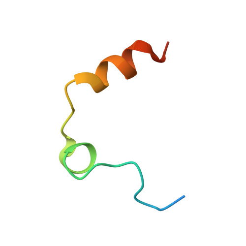
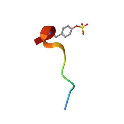
  • Primary Citation Related Structures: 
    1W7H, 1WAX, 1WAY, 1WBG, 1WBO, 1WBU, 1WCC

  • PubMed Abstract: 

    Fragment screening offers an alternative to traditional screening for discovering new leads in drug discovery programs. This paper describes a fragment screening methodology based on high throughput X-ray crystallography. The method is illustrated against five proteins (p38 MAP kinase, CDK2, thrombin, ribonuclease A, and PTP1B). The fragments identified have weak potency (>100 microM) but are efficient binders relative to their size and may therefore represent suitable starting points for evolution to good quality lead compounds. The examples illustrate that a range of molecular interactions (i.e., lipophilic, charge-charge, neutral hydrogen bonds) can drive fragment binding and also that fragments can induce protein movement. We believe that the method has great potential for the discovery of novel lead compounds against a range of targets, and the companion paper illustrates how lead compounds have been identified for p38 MAP kinase starting from fragments such as those described in this paper.


  • Organizational Affiliation
    • Astex Technology, 436 Cambridge Science Park, Milton Road, Cambridge, CB4 0QA, United Kingdom.

Macromolecule Content 

  • Total Structure Weight: 35.74 kDa 
  • Atom Count: 2,777 
  • Modeled Residue Count: 290 
  • Deposited Residue Count: 305 
  • Unique protein chains: 3

Macromolecules

Find similar proteins by:|  3D Structure
Entity ID: 1
MoleculeChains  Sequence LengthOrganismDetailsImage
THROMBIN LIGHT CHAIN36Homo sapiensMutation(s): 0 
EC: 3.4.21.5
UniProt & NIH Common Fund Data Resources
Find proteins for P00734 (Homo sapiens)
Explore P00734 
Go to UniProtKB:  P00734
PHAROS:  P00734
GTEx:  ENSG00000180210 
Entity Groups
Sequence Clusters30% Identity50% Identity70% Identity90% Identity95% Identity100% Identity
UniProt GroupP00734
Sequence Annotations
Expand
Reference Sequence
Find similar proteins by:|  3D Structure
Entity ID: 2
MoleculeChains  Sequence LengthOrganismDetailsImage
THROMBIN HEAVY CHAIN259Homo sapiensMutation(s): 0 
EC: 3.4.21.5
UniProt & NIH Common Fund Data Resources
Find proteins for P00734 (Homo sapiens)
Explore P00734 
Go to UniProtKB:  P00734
PHAROS:  P00734
GTEx:  ENSG00000180210 
Entity Groups
Sequence Clusters30% Identity50% Identity70% Identity90% Identity95% Identity100% Identity
UniProt GroupP00734
Sequence Annotations
Expand
Reference Sequence
Find similar proteins by:  Sequence   |   3D Structure  
Entity ID: 3
MoleculeChains  Sequence LengthOrganismDetailsImage
HIRUGENC [auth I]10Hirudo medicinalisMutation(s): 0 
UniProt
Find proteins for P01050 (Hirudo medicinalis)
Explore P01050 
Go to UniProtKB:  P01050
Entity Groups
Sequence Clusters30% Identity50% Identity70% Identity90% Identity95% Identity100% Identity
UniProt GroupP01050
Sequence Annotations
Expand
Reference Sequence

Small Molecules

Modified Residues  1 Unique
IDChains TypeFormula2D DiagramParent
TYS
Query on TYS
C [auth I]L-PEPTIDE LINKINGC9 H11 N O6 STYR

Experimental Data & Validation

Experimental Data

  • Method: X-RAY DIFFRACTION
  • Resolution: 2.02 Å
  • R-Value Free:  0.231 (Depositor) 
  • R-Value Work:  0.158 (Depositor) 
  • R-Value Observed: 0.161 (Depositor) 
Space Group: C 1 2 1
Unit Cell:
Length ( Å )Angle ( ˚ )
a = 70.503α = 90
b = 71.512β = 100.46
c = 72.205γ = 90
Software Package:
Software NamePurpose
REFMACrefinement
d*TREKdata reduction
d*TREKdata scaling
IN-HOUSEphasing

Structure Validation

View Full Validation Report


Entry History 

Deposition Data

Revision History  (Full details and data files)

  • Version 1.0: 2005-01-27
    Type: Initial release
  • Version 1.1: 2011-07-13
    Changes: Atomic model, Database references, Derived calculations, Non-polymer description, Structure summary, Version format compliance
  • Version 1.2: 2017-06-28
    Changes: Data collection
  • Version 1.3: 2023-12-13
    Changes: Data collection, Database references, Derived calculations, Refinement description
  • Version 1.4: 2024-11-06
    Changes: Structure summary