5YQ5

Crystal structure of human osteomodulin


Experimental Data Snapshot

  • Method: X-RAY DIFFRACTION
  • Resolution: 2.17 Å
  • R-Value Free: 0.245 
  • R-Value Work: 0.217 
  • R-Value Observed: 0.218 

wwPDB Validation   3D Report Full Report


Ligand Structure Quality Assessment 


This is version 1.3 of the entry. See complete history


Literature

Molecular basis for governing the morphology of type-I collagen fibrils by Osteomodulin.

Tashima, T.Nagatoishi, S.Caaveiro, J.M.M.Nakakido, M.Sagara, H.Kusano-Arai, O.Iwanari, H.Mimuro, H.Hamakubo, T.Ohnuma, S.I.Tsumoto, K.

(2018) Commun Biol 1: 33-33

  • DOI: https://doi.org/10.1038/s42003-018-0038-2
  • Primary Citation of Related Structures:  
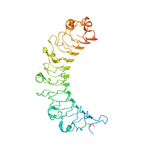
    5YQ5

  • PubMed Abstract: 

    Small leucine-rich repeat proteoglycan (SLRP) proteins have an important role in the organization of the extracellular matrix, especially in the formation of collagen fibrils. However, the mechanism governing the shape of collagen fibrils is poorly understood. Here, we report that the protein Osteomodulin (OMD) of the SLRP family is a monomeric protein in solution that interacts with type-I collagen. This interaction is dominated by weak electrostatic forces employing negatively charged residues of OMD, in particular Glu284 and Glu303, and controlled by entropic factors. The protein OMD establishes a fast-binding equilibrium with collagen, where OMD may engage not only with individual collagen molecules, but also with the growing fibrils. This weak electrostatic interaction is carefully balanced so it modulates the shape of the fibrils without compromising their viability.


  • Organizational Affiliation

    Department of Chemistry & Biotechnology, School of Engineering, The University of Tokyo, Tokyo, 108-8639, Japan.


Macromolecules
Find similar proteins by:  (by identity cutoff)  |  3D Structure
Entity ID: 1
MoleculeChains Sequence LengthOrganismDetailsImage
Osteomodulin
A, B, C, D
430Homo sapiensMutation(s): 0 
Gene Names: OMDSLRR2CUNQ190/PRO216
UniProt & NIH Common Fund Data Resources
Find proteins for Q99983 (Homo sapiens)
Explore Q99983 
Go to UniProtKB:  Q99983
PHAROS:  Q99983
GTEx:  ENSG00000127083 
Entity Groups  
Sequence Clusters30% Identity50% Identity70% Identity90% Identity95% Identity100% Identity
UniProt GroupQ99983
Sequence Annotations
Expand
  • Reference Sequence
Small Molecules
Ligands 2 Unique
IDChains Name / Formula / InChI Key2D Diagram3D Interactions
NAG
Query on NAG

Download Ideal Coordinates CCD File 
E [auth A]
F [auth A]
G [auth A]
H [auth A]
I [auth B]
E [auth A],
F [auth A],
G [auth A],
H [auth A],
I [auth B],
J [auth B],
K [auth B],
M [auth C],
N [auth C],
O [auth C],
P [auth D],
Q [auth D],
R [auth D]
2-acetamido-2-deoxy-beta-D-glucopyranose
C8 H15 N O6
OVRNDRQMDRJTHS-FMDGEEDCSA-N
PO4
Query on PO4

Download Ideal Coordinates CCD File 
L [auth B],
S [auth D]
PHOSPHATE ION
O4 P
NBIIXXVUZAFLBC-UHFFFAOYSA-K
Experimental Data & Validation

Experimental Data

  • Method: X-RAY DIFFRACTION
  • Resolution: 2.17 Å
  • R-Value Free: 0.245 
  • R-Value Work: 0.217 
  • R-Value Observed: 0.218 
  • Space Group: P 1 21 1
Unit Cell:
Length ( Å )Angle ( ˚ )
a = 75.64α = 90
b = 110.7β = 106.97
c = 122.15γ = 90
Software Package:
Software NamePurpose
REFMACrefinement
iMOSFLMdata reduction
SCALAdata scaling
PHASERphasing

Structure Validation

View Full Validation Report


Ligand Structure Quality Assessment 


Entry History & Funding Information

Deposition Data


Funding OrganizationLocationGrant Number
JSPSJapan16H02420
JSPSJapan15K06962

Revision History  (Full details and data files)

  • Version 1.0: 2018-04-25
    Type: Initial release
  • Version 1.1: 2018-10-17
    Changes: Data collection, Database references
  • Version 1.2: 2020-07-29
    Type: Remediation
    Reason: Carbohydrate remediation
    Changes: Data collection, Derived calculations, Structure summary
  • Version 1.3: 2023-11-22
    Changes: Data collection, Database references, Refinement description, Structure summary