5OYA

Unusual posttranslational modifications revealed in crystal structures of diatom Rubisco.


Experimental Data Snapshot

  • Method: X-RAY DIFFRACTION
  • Resolution: 1.80 Å
  • R-Value Free: 0.192 
  • R-Value Work: 0.167 
  • R-Value Observed: 0.168 

wwPDB Validation   3D Report Full Report


Ligand Structure Quality Assessment 


This is version 1.4 of the entry. See complete history


Literature

Structural and functional analyses of Rubisco from arctic diatom species reveal unusual posttranslational modifications.

Valegard, K.Andralojc, P.J.Haslam, R.P.Pearce, F.G.Eriksen, G.K.Madgwick, P.J.Kristoffersen, A.K.van Lun, M.Klein, U.Eilertsen, H.C.Parry, M.A.J.Andersson, I.

(2018) J Biol Chem 293: 13033-13043

  • DOI: https://doi.org/10.1074/jbc.RA118.003518
  • Primary Citation of Related Structures:  
    5MZ2, 5N9Z, 5OYA, 6FTL

  • PubMed Abstract: 

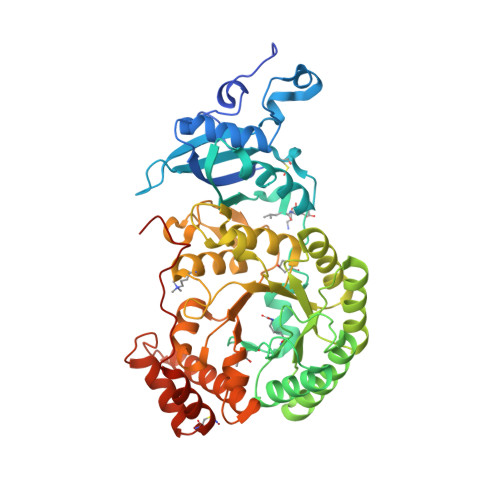
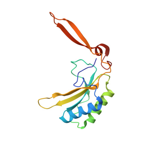
    The catalytic performance of the major CO 2 -assimilating enzyme, ribulose-1,5-bisphosphate carboxylase/oxygenase (Rubisco), restricts photosynthetic productivity. Natural diversity in the catalytic properties of Rubisco indicates possibilities for improvement. Oceanic phytoplankton contain some of the most efficient Rubisco enzymes, and diatoms in particular are responsible for a significant proportion of total marine primary production as well as being a major source of CO 2 sequestration in polar cold waters. Until now, the biochemical properties and three-dimensional structures of Rubisco from diatoms were unknown. Here, diatoms from arctic waters were collected, cultivated, and analyzed for their CO 2 -fixing capability. We characterized the kinetic properties of five and determined the crystal structures of four Rubiscos selected for their high CO 2 -fixing efficiency. The DNA sequences of the rbc L and rbc S genes of the selected diatoms were similar, reflecting their close phylogenetic relationship. The V max and K m for the oxygenase and carboxylase activities at 25 °C and the specificity factors ( S c/o ) at 15, 25, and 35 °C were determined. The S c/o values were high, approaching those of mono- and dicot plants, thus exhibiting good selectivity for CO 2 relative to O 2 Structurally, diatom Rubiscos belong to form I C/D, containing small subunits characterized by a short βA-βB loop and a C-terminal extension that forms a β-hairpin structure (βE-βF loop). Of note, the diatom Rubiscos featured a number of posttranslational modifications of the large subunit, including 4-hydroxyproline, β-hydroxyleucine, hydroxylated and nitrosylated cysteine, mono- and dihydroxylated lysine, and trimethylated lysine. Our studies suggest adaptation toward achieving efficient CO 2 fixation in arctic diatom Rubiscos.


  • Organizational Affiliation

    From the Department of Cell and Molecular Biology, Uppsala University, Box 596, S-751 24 Uppsala, Sweden.


Macromolecules
Find similar proteins by:  (by identity cutoff)  |  3D Structure
Entity ID: 1
MoleculeChains Sequence LengthOrganismDetailsImage
Rubisco large subunit
A, B, D, E, F
A, B, D, E, F, G, H
490Chaetoceros socialisMutation(s): 0 
UniProt
Find proteins for A0A3B6UEF4 (Chaetoceros socialis)
Explore A0A3B6UEF4 
Go to UniProtKB:  A0A3B6UEF4
Entity Groups  
Sequence Clusters30% Identity50% Identity70% Identity90% Identity95% Identity100% Identity
UniProt GroupA0A3B6UEF4
Sequence Annotations
Expand
  • Reference Sequence
Find similar proteins by:  (by identity cutoff)  |  3D Structure
Entity ID: 2
MoleculeChains Sequence LengthOrganismDetailsImage
Rubisco large subunit490Chaetoceros socialisMutation(s): 0 
UniProt
Find proteins for A0A3B6UEF4 (Chaetoceros socialis)
Explore A0A3B6UEF4 
Go to UniProtKB:  A0A3B6UEF4
Entity Groups  
Sequence Clusters30% Identity50% Identity70% Identity90% Identity95% Identity100% Identity
UniProt GroupA0A3B6UEF4
Sequence Annotations
Expand
  • Reference Sequence
Find similar proteins by:  (by identity cutoff)  |  3D Structure
Entity ID: 3
MoleculeChains Sequence LengthOrganismDetailsImage
Rubisco small subunit
I, J, K, L, M
I, J, K, L, M, N, O, P
139Chaetoceros socialisMutation(s): 0 
UniProt
Find proteins for A0A3B6UEF5 (Chaetoceros socialis)
Explore A0A3B6UEF5 
Go to UniProtKB:  A0A3B6UEF5
Entity Groups  
Sequence Clusters30% Identity50% Identity70% Identity90% Identity95% Identity100% Identity
UniProt GroupA0A3B6UEF5
Sequence Annotations
Expand
  • Reference Sequence
Small Molecules
Ligands 3 Unique
IDChains Name / Formula / InChI Key2D Diagram3D Interactions
CAP
Query on CAP

Download Ideal Coordinates CCD File 
BA [auth F]
DA [auth G]
FA [auth H]
R [auth A]
T [auth B]
BA [auth F],
DA [auth G],
FA [auth H],
R [auth A],
T [auth B],
V [auth C],
X [auth D],
Z [auth E]
2-CARBOXYARABINITOL-1,5-DIPHOSPHATE
C6 H14 O13 P2
ITHCSGCUQDMYAI-ZMIZWQJLSA-N
EDO
Query on EDO

Download Ideal Coordinates CCD File 
AB [auth O]
BB [auth O]
CB [auth O]
DB [auth P]
EB [auth P]
AB [auth O],
BB [auth O],
CB [auth O],
DB [auth P],
EB [auth P],
FB [auth P],
GA [auth I],
HA [auth I],
IA [auth I],
KA [auth J],
LA [auth J],
MA [auth J],
NA [auth K],
OA [auth K],
PA [auth K],
QA [auth L],
RA [auth L],
SA [auth L],
TA [auth M],
UA [auth M],
VA [auth M],
XA [auth N],
YA [auth N],
ZA [auth N]
1,2-ETHANEDIOL
C2 H6 O2
LYCAIKOWRPUZTN-UHFFFAOYSA-N
MG
Query on MG

Download Ideal Coordinates CCD File 
AA [auth F]
CA [auth G]
EA [auth H]
JA [auth J]
Q [auth A]
AA [auth F],
CA [auth G],
EA [auth H],
JA [auth J],
Q [auth A],
S [auth B],
U [auth C],
W [auth D],
WA [auth N],
Y [auth E]
MAGNESIUM ION
Mg
JLVVSXFLKOJNIY-UHFFFAOYSA-N
Modified Residues  6 Unique
IDChains TypeFormula2D DiagramParent
CSO
Query on CSO
A, B, D, E, F
A, B, D, E, F, G, H
L-PEPTIDE LINKINGC3 H7 N O3 SCYS
HL2
Query on HL2
A, B, D, E, F
A, B, D, E, F, G, H
L-PEPTIDE LINKINGC6 H13 N O3LEU
HYP
Query on HYP
A, B, D, E, F
A, B, D, E, F, G, H
L-PEPTIDE LINKINGC5 H9 N O3PRO
KCX
Query on KCX
A, B, D, E, F
A, B, D, E, F, G, H
L-PEPTIDE LINKINGC7 H14 N2 O4LYS
M3L
Query on M3L
A, B, D, E, F
A, B, D, E, F, G, H
L-PEPTIDE LINKINGC9 H21 N2 O2LYS
SNC
Query on SNC
A, B, D, E, F
A, B, D, E, F, G, H
L-PEPTIDE LINKINGC3 H6 N2 O3 SCYS
Experimental Data & Validation

Experimental Data

  • Method: X-RAY DIFFRACTION
  • Resolution: 1.80 Å
  • R-Value Free: 0.192 
  • R-Value Work: 0.167 
  • R-Value Observed: 0.168 
  • Space Group: C 1 2 1
Unit Cell:
Length ( Å )Angle ( ˚ )
a = 118.17α = 90
b = 219.11β = 90.19
c = 220.23γ = 90
Software Package:
Software NamePurpose
REFMACrefinement
XDSdata processing
XSCALEdata scaling
PHASERphasing
Omodel building

Structure Validation

View Full Validation Report


Ligand Structure Quality Assessment 


Entry History & Funding Information

Deposition Data


Funding OrganizationLocationGrant Number
European UnionSwedenQLK3-CT-2002-01945

Revision History  (Full details and data files)

  • Version 1.0: 2018-06-27
    Type: Initial release
  • Version 1.1: 2018-07-04
    Changes: Data collection, Database references
  • Version 1.2: 2018-09-05
    Changes: Data collection, Database references
  • Version 1.3: 2019-04-17
    Changes: Data collection, Other, Structure summary
  • Version 1.4: 2024-01-17
    Changes: Data collection, Database references, Derived calculations, Refinement description