4S1K

Structure of Uranotaenia sapphirina cypovirus (CPV17) polyhedrin at 100 K


Experimental Data Snapshot

  • Method: X-RAY DIFFRACTION
  • Resolution: 2.20 Å
  • R-Value Free: 0.199 
  • R-Value Work: 0.148 
  • R-Value Observed: 0.151 

wwPDB Validation   3D Report Full Report


Ligand Structure Quality Assessment 


This is version 1.1 of the entry. See complete history


Literature

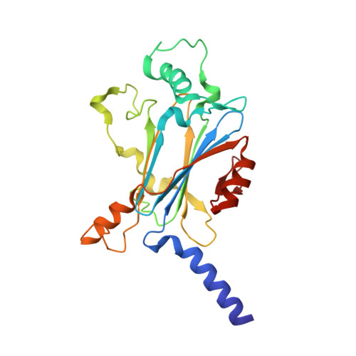
Structure of CPV17 polyhedrin determined by the improved analysis of serial femtosecond crystallographic data.

Ginn, H.M.Messerschmidt, M.Ji, X.Zhang, H.Axford, D.Gildea, R.J.Winter, G.Brewster, A.S.Hattne, J.Wagner, A.Grimes, J.M.Evans, G.Sauter, N.K.Sutton, G.Stuart, D.I.

(2015) Nat Commun 6: 6435-6435

  • DOI: https://doi.org/10.1038/ncomms7435
  • Primary Citation of Related Structures:  
    4S1K, 4S1L

  • PubMed Abstract: 

    The X-ray free-electron laser (XFEL) allows the analysis of small weakly diffracting protein crystals, but has required very many crystals to obtain good data. Here we use an XFEL to determine the room temperature atomic structure for the smallest cytoplasmic polyhedrosis virus polyhedra yet characterized, which we failed to solve at a synchrotron. These protein microcrystals, roughly a micron across, accrue within infected cells. We use a new physical model for XFEL diffraction, which better estimates the experimental signal, delivering a high-resolution XFEL structure (1.75 Å), using fewer crystals than previously required for this resolution. The crystal lattice and protein core are conserved compared with a polyhedrin with less than 10% sequence identity. We explain how the conserved biological phenotype, the crystal lattice, is maintained in the face of extreme environmental challenge and massive evolutionary divergence. Our improved methods should open up more challenging biological samples to XFEL analysis.


  • Organizational Affiliation

    Division of Structural Biology, The Wellcome Trust Centre for Human Genetics, University of Oxford, Roosevelt Drive, Oxford, Oxfordshire OX3 7BN, UK.


Macromolecules
Find similar proteins by:  (by identity cutoff)  |  3D Structure
Entity ID: 1
MoleculeChains Sequence LengthOrganismDetailsImage
Polyhedrin237Uranotaenia sapphirina cypovirusMutation(s): 0 
UniProt
Find proteins for Q5EK29 (Uranotaenia sapphirina cypovirus)
Explore Q5EK29 
Go to UniProtKB:  Q5EK29
Entity Groups  
Sequence Clusters30% Identity50% Identity70% Identity90% Identity95% Identity100% Identity
UniProt GroupQ5EK29
Sequence Annotations
Expand
  • Reference Sequence
Small Molecules
Ligands 2 Unique
IDChains Name / Formula / InChI Key2D Diagram3D Interactions
ATP
Query on ATP

Download Ideal Coordinates CCD File 
B [auth A]ADENOSINE-5'-TRIPHOSPHATE
C10 H16 N5 O13 P3
ZKHQWZAMYRWXGA-KQYNXXCUSA-N
MG
Query on MG

Download Ideal Coordinates CCD File 
C [auth A]MAGNESIUM ION
Mg
JLVVSXFLKOJNIY-UHFFFAOYSA-N
Experimental Data & Validation

Experimental Data

  • Method: X-RAY DIFFRACTION
  • Resolution: 2.20 Å
  • R-Value Free: 0.199 
  • R-Value Work: 0.148 
  • R-Value Observed: 0.151 
  • Space Group: I 2 3
Unit Cell:
Length ( Å )Angle ( ˚ )
a = 104.879α = 90
b = 104.879β = 90
c = 104.879γ = 90
Software Package:
Software NamePurpose
DIALSdata collection
PHASERphasing
REFMACrefinement
DIALSdata reduction
DIALSdata scaling

Structure Validation

View Full Validation Report


Ligand Structure Quality Assessment 


Entry History 

Revision History  (Full details and data files)

  • Version 1.0: 2015-03-25
    Type: Initial release
  • Version 1.1: 2024-02-28
    Changes: Data collection, Database references, Derived calculations