4OIK

(Quasi-)Racemic X-ray crystal structure of glycosylated chemokine Ser-CCL1.


Experimental Data Snapshot

  • Method: X-RAY DIFFRACTION
  • Resolution: 2.10 Å
  • R-Value Free: 0.276 
  • R-Value Work: 0.229 
  • R-Value Observed: 0.231 

wwPDB Validation   3D Report Full Report


Ligand Structure Quality Assessment 


This is version 1.4 of the entry. See complete history


Literature

(Quasi-)Racemic X-ray Structures of Glycosylated and Non-Glycosylated Forms of the Chemokine Ser-CCL1 Prepared by Total Chemical Synthesis.

Okamoto, R.Mandal, K.Sawaya, M.R.Kajihara, Y.Yeates, T.O.Kent, S.B.

(2014) Angew Chem Int Ed Engl 53: 5194-5198

  • DOI: https://doi.org/10.1002/anie.201400679
  • Primary Citation of Related Structures:  
    4OIJ, 4OIK

  • PubMed Abstract: 

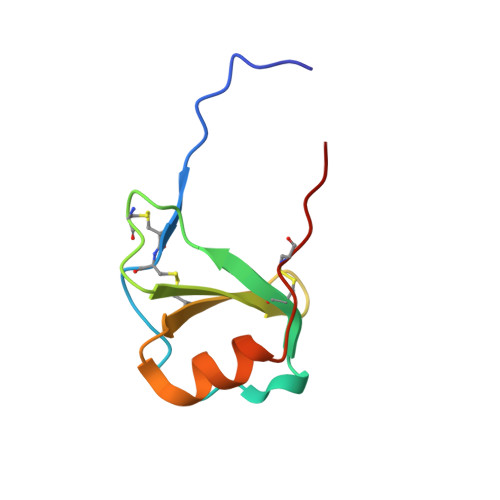
    Our goal was to obtain the X-ray crystal structure of the glycosylated chemokine Ser-CCL1. Glycoproteins can be hard to crystallize because of the heterogeneity of the oligosaccharide (glycan) moiety. We used glycosylated Ser-CCL1 that had been prepared by total chemical synthesis as a homogeneous compound containing an N-linked asialo biantennary nonasaccharide glycan moiety of defined covalent structure. Facile crystal formation occurred from a quasi-racemic mixture consisting of glycosylated L-protein and non-glycosylated-D-protein, while no crystals were obtained from the glycosylated L-protein alone. The structure was solved at a resolution of 2.6-2.1 Å. However, the glycan moiety was disordered: only the N-linked GlcNAc sugar was well-defined in the electron density map. A racemic mixture of the protein enantiomers L-Ser-CCL1 and D-Ser-CCL1 was also crystallized, and the structure of the true racemate was solved at a resolution of 2.7-2.15 Å. Superimposition of the structures of the protein moieties of L-Ser-CCL1 and glycosylated-L-Ser-CCL1 revealed there was no significant alteration of the protein structure by N-glycosylation.


  • Organizational Affiliation

    Departments of Chemistry: Biochemistry & Molecular Biology, Institute for Biophysical Dynamics, University of Chicago, Chicago, IL 60637 (USA); Current address: Department of Chemistry, Graduate School of Science, Osaka University, Toyonaka, Osaka, 560-0043, JAPAN.. skent@uchicago.edu, rokamoto@chem.sci.osaka-u.ac.jp, rokamoto@chem.sci.osaka-u.ac.jp.


Macromolecules
Find similar proteins by:  (by identity cutoff)  |  3D Structure
Entity ID: 1
MoleculeChains Sequence LengthOrganismDetailsImage
C-C motif chemokine 1
A, B
74Homo sapiensMutation(s): 0 
UniProt & NIH Common Fund Data Resources
Find proteins for P22362 (Homo sapiens)
Explore P22362 
Go to UniProtKB:  P22362
PHAROS:  P22362
GTEx:  ENSG00000108702 
Entity Groups  
Sequence Clusters30% Identity50% Identity70% Identity90% Identity95% Identity100% Identity
UniProt GroupP22362
Sequence Annotations
Expand
  • Reference Sequence
Find similar proteins by:  (by identity cutoff)  |  3D Structure
Entity ID: 2
MoleculeChains Sequence LengthOrganismDetailsImage
D-Ser-CCL1
C, D
74synthetic constructMutation(s): 0 
Sequence Annotations
Expand
  • Reference Sequence
Small Molecules
Ligands 3 Unique
IDChains Name / Formula / InChI Key2D Diagram3D Interactions
NAG
Query on NAG

Download Ideal Coordinates CCD File 
E [auth A],
N [auth B]
2-acetamido-2-deoxy-beta-D-glucopyranose
C8 H15 N O6
OVRNDRQMDRJTHS-FMDGEEDCSA-N
CIT
Query on CIT

Download Ideal Coordinates CCD File 
CA [auth C],
IA [auth D]
CITRIC ACID
C6 H8 O7
KRKNYBCHXYNGOX-UHFFFAOYSA-N
SO4
Query on SO4

Download Ideal Coordinates CCD File 
AA [auth C]
BA [auth C]
DA [auth D]
EA [auth D]
F [auth A]
AA [auth C],
BA [auth C],
DA [auth D],
EA [auth D],
F [auth A],
FA [auth D],
G [auth A],
GA [auth D],
H [auth A],
HA [auth D],
I [auth A],
J [auth A],
K [auth A],
L [auth A],
M [auth A],
O [auth B],
P [auth B],
Q [auth B],
R [auth B],
S [auth B],
T [auth B],
U [auth B],
V [auth C],
W [auth C],
X [auth C],
Y [auth C],
Z [auth C]
SULFATE ION
O4 S
QAOWNCQODCNURD-UHFFFAOYSA-L
Experimental Data & Validation

Experimental Data

  • Method: X-RAY DIFFRACTION
  • Resolution: 2.10 Å
  • R-Value Free: 0.276 
  • R-Value Work: 0.229 
  • R-Value Observed: 0.231 
  • Space Group: P 1
Unit Cell:
Length ( Å )Angle ( ˚ )
a = 45.41α = 111.23
b = 51.68β = 106.33
c = 51.33γ = 108.68
Software Package:
Software NamePurpose
PHASERphasing
BUSTERrefinement
HKL-2000data reduction
SCALEPACKdata scaling

Structure Validation

View Full Validation Report


Ligand Structure Quality Assessment 


Entry History 

Deposition Data

Revision History  (Full details and data files)

  • Version 1.0: 2014-05-07
    Type: Initial release
  • Version 1.1: 2014-05-21
    Changes: Database references
  • Version 1.2: 2020-07-29
    Type: Remediation
    Reason: Carbohydrate remediation
    Changes: Data collection, Derived calculations, Structure summary
  • Version 1.3: 2023-09-20
    Changes: Data collection, Database references, Refinement description, Structure summary
  • Version 1.4: 2023-12-06
    Changes: Data collection