3H5C

X-Ray Structure of Protein Z-Protein Z Inhibitor Complex


Experimental Data Snapshot

  • Method: X-RAY DIFFRACTION
  • Resolution: 3.26 Å
  • R-Value Free: 0.269 
  • R-Value Work: 0.201 
  • R-Value Observed: 0.206 

wwPDB Validation   3D Report Full Report


Ligand Structure Quality Assessment 


This is version 1.3 of the entry. See complete history


Literature

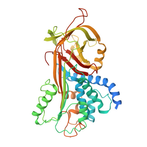
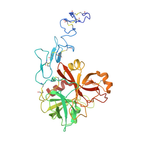
Basis for the specificity and activation of the serpin protein Z-dependent proteinase inhibitor (ZPI) as an inhibitor of membrane-associated factor Xa.

Huang, X.Dementiev, A.Olson, S.T.Gettins, P.G.

(2010) J Biol Chem 285: 20399-20409

  • DOI: https://doi.org/10.1074/jbc.M110.112748
  • Primary Citation of Related Structures:  
    3H5C

  • PubMed Abstract: 

    The serpin ZPI is a protein Z (PZ)-dependent specific inhibitor of membrane-associated factor Xa (fXa) despite having an unfavorable P1 Tyr. PZ accelerates the inhibition reaction approximately 2000-fold in the presence of phospholipid and Ca(2+). To elucidate the role of PZ, we determined the x-ray structure of Gla-domainless PZ (PZ(DeltaGD)) complexed with protein Z-dependent proteinase inhibitor (ZPI). The PZ pseudocatalytic domain bound ZPI at a novel site through ionic and polar interactions. Mutation of four ZPI contact residues eliminated PZ binding and membrane-dependent PZ acceleration of fXa inhibition. Modeling of the ternary Michaelis complex implicated ZPI residues Glu-313 and Glu-383 in fXa binding. Mutagenesis established that only Glu-313 is important, contributing approximately 5-10-fold to rate acceleration of fXa and fXIa inhibition. Limited conformational change in ZPI resulted from PZ binding, which contributed only approximately 2-fold to rate enhancement. Instead, template bridging from membrane association, together with previously demonstrated interaction of the fXa and ZPI Gla domains, resulted in an additional approximately 1000-fold rate enhancement. To understand why ZPI has P1 tyrosine, we examined a P1 Arg variant. This reacted at a diffusion-limited rate with fXa, even without PZ, and predominantly as substrate, reflecting both rapid acylation and deacylation. P1 tyrosine thus ensures that reaction with fXa or most other arginine-specific proteinases is insignificant unless PZ binds and localizes ZPI and fXa on the membrane, where the combined effects of Gla-Gla interaction, template bridging, and interaction of fXa with Glu-313 overcome the unfavorability of P1 Tyr and ensure a high rate of reaction as an inhibitor.


  • Organizational Affiliation

    Center for Molecular Biology of Oral Diseases, University of Illinois at Chicago, Chicago, Illinois 60607, USA.


Macromolecules
Find similar proteins by:  (by identity cutoff)  |  3D Structure
Entity ID: 1
MoleculeChains Sequence LengthOrganismDetailsImage
Protein Z-dependent protease inhibitor423Homo sapiensMutation(s): 0 
Gene Names: SERPINA10ZPIUNQ707/PRO1358
UniProt & NIH Common Fund Data Resources
Find proteins for Q9UK55 (Homo sapiens)
Explore Q9UK55 
Go to UniProtKB:  Q9UK55
PHAROS:  Q9UK55
GTEx:  ENSG00000140093 
Entity Groups  
Sequence Clusters30% Identity50% Identity70% Identity90% Identity95% Identity100% Identity
UniProt GroupQ9UK55
Sequence Annotations
Expand
  • Reference Sequence
Find similar proteins by:  (by identity cutoff)  |  3D Structure
Entity ID: 2
MoleculeChains Sequence LengthOrganismDetailsImage
Vitamin K-dependent protein Z317Homo sapiensMutation(s): 0 
UniProt & NIH Common Fund Data Resources
Find proteins for P22891 (Homo sapiens)
Explore P22891 
Go to UniProtKB:  P22891
PHAROS:  P22891
GTEx:  ENSG00000126231 
Entity Groups  
Sequence Clusters30% Identity50% Identity70% Identity90% Identity95% Identity100% Identity
UniProt GroupP22891
Sequence Annotations
Expand
  • Reference Sequence
Experimental Data & Validation

Experimental Data

  • Method: X-RAY DIFFRACTION
  • Resolution: 3.26 Å
  • R-Value Free: 0.269 
  • R-Value Work: 0.201 
  • R-Value Observed: 0.206 
  • Space Group: P 65 2 2
Unit Cell:
Length ( Å )Angle ( ˚ )
a = 119.934α = 90
b = 119.934β = 90
c = 347.672γ = 120
Software Package:
Software NamePurpose
XDSdata scaling
PHASERphasing
REFMACrefinement
XDSdata reduction

Structure Validation

View Full Validation Report


Ligand Structure Quality Assessment 


Entry History 

Deposition Data

Revision History  (Full details and data files)

  • Version 1.0: 2010-04-28
    Type: Initial release
  • Version 1.1: 2011-07-13
    Changes: Version format compliance
  • Version 1.2: 2017-11-01
    Changes: Refinement description
  • Version 1.3: 2020-07-29
    Type: Remediation
    Reason: Carbohydrate remediation
    Changes: Data collection, Derived calculations, Structure summary