2QTY

Crystal Structure of mouse ADP-ribosylhydrolase 3 (mARH3)


Experimental Data Snapshot

  • Method: X-RAY DIFFRACTION
  • Resolution: 1.80 Å
  • R-Value Free: 0.198 
  • R-Value Work: 0.167 
  • R-Value Observed: 0.168 

wwPDB Validation   3D Report Full Report


This is version 1.2 of the entry. See complete history


Literature

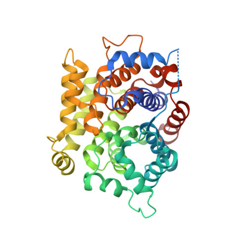
Structure of mouse ADP-ribosylhydrolase 3 (mARH3).

Mueller-Dieckmann, C.Kernstock, S.Mueller-Dieckmann, J.Weiss, M.S.Koch-Nolte, F.

(2008) Acta Crystallogr Sect F Struct Biol Cryst Commun 64: 156-162

  • DOI: https://doi.org/10.1107/S1744309108001413
  • Primary Citation of Related Structures:  
    2QTY

  • PubMed Abstract: 

    ADP-ribosylation is a reversible and covalent post-translational modification in which the attachment of ADP-ribose is catalyzed by ADP-ribosyltransferases and the removal of ADP-ribose is catalyzed by ADP-ribosylhydrolases. ADP-ribosylhydrolase 3 from mouse, consisting of 347 amino-acid residues, has been cloned, purified and crystallized. The three-dimensional structure has been resolved at a resolution of 1.8 A. The structure constitutes a compact all-alpha-helical protein with two Mg(2+) ions located in the active-site crevice. A structural comparison of mouse ADP-ribosylhydrolase 3 with its human orthologue shows a high degree of structural similarity. Furthermore, four prokaryotic proteins deposited in the PDB could be identified as being structurally related.


  • Organizational Affiliation

    ESRF, 6 Rue Jules Horowitz, F-38043 Grenoble CEDEX 09, France. muellerd@esrf.fr


Macromolecules
Find similar proteins by:  (by identity cutoff)  |  3D Structure
Entity ID: 1
MoleculeChains Sequence LengthOrganismDetailsImage
Poly(ADP-ribose) glycohydrolase ARH3
A, B
347Mus musculusMutation(s): 0 
EC: 3.2.1.143
UniProt & NIH Common Fund Data Resources
Find proteins for Q8CG72 (Mus musculus)
Explore Q8CG72 
Go to UniProtKB:  Q8CG72
IMPC:  MGI:2140364
Entity Groups  
Sequence Clusters30% Identity50% Identity70% Identity90% Identity95% Identity100% Identity
UniProt GroupQ8CG72
Sequence Annotations
Expand
  • Reference Sequence
Experimental Data & Validation

Experimental Data

  • Method: X-RAY DIFFRACTION
  • Resolution: 1.80 Å
  • R-Value Free: 0.198 
  • R-Value Work: 0.167 
  • R-Value Observed: 0.168 
  • Space Group: P 1 21 1
Unit Cell:
Length ( Å )Angle ( ˚ )
a = 53.877α = 90
b = 60.096β = 90.09
c = 91.782γ = 90
Software Package:
Software NamePurpose
REFMACrefinement
PDB_EXTRACTdata extraction
HKL-2000data collection
DENZOdata reduction
SCALEPACKdata scaling
MOLREPphasing

Structure Validation

View Full Validation Report


Entry History 

Deposition Data

Revision History  (Full details and data files)

  • Version 1.0: 2008-03-04
    Type: Initial release
  • Version 1.1: 2011-07-13
    Changes: Advisory, Version format compliance
  • Version 1.2: 2023-08-30
    Changes: Data collection, Database references, Derived calculations, Refinement description