9GMU | pdb_00009gmu

Structure ofhuman aM ligand binding domain in complex with the aCR3 nanobody


Experimental Data Snapshot

  • Method: X-RAY DIFFRACTION
  • Resolution: 3.80 Å
  • R-Value Free: 
    0.298 (Depositor), 0.300 (DCC) 
  • R-Value Work: 
    0.242 (Depositor), 0.241 (DCC) 
  • R-Value Observed: 
    0.245 (Depositor) 

Starting Model: experimental
View more details

wwPDB Validation 3D Report Full Report

Validation slider image for 9GMU

This is version 1.1 of the entry. See complete history

Literature

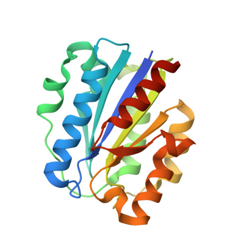
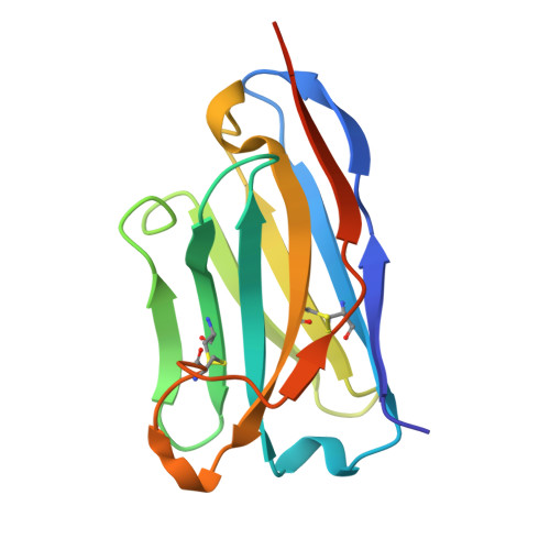
Three cryo-EM structures of complement C3d-bound alpha M beta 2 reveal an unexpected layer of dynamics for alpha I-containing integrin receptors.

Lorentzen, J.Fruergaard, M.U.Lukacsi, S.Jorgensen, M.H.van Veghel, T.L.G.Jensen, R.K.Pietrzak-Lichwa, K.J.Bajtay, Z.Horejsi, V.Flygaard, R.K.Vorselen, D.Mortensen, S.A.Andersen, G.R.

(2026) Sci Adv 12: eaea7241-eaea7241

  • DOI: https://doi.org/10.1126/sciadv.aea7241
  • Primary Citation Related Structures: 
    9GMU, 9HLL, 9RM9, 9RMA, 9T3Y, 9T5V, 9T5W, 9T5Z

  • PubMed Abstract: 

    Integrins are heterodimeric membrane proteins acting as mechanosensing receptors. Nine human α-subunits contain a ligand binding αI domain, but how ligands activate αI integrins are not understood. We present cryo-EM structures of the αI integrin α M β 2 in complex with the C3d ligand. The ligand-bound αI domain appears to have two major opposite orientations relative to the β 2 subunit. Ligand binding induces an ordered conformation of the α M internal ligand region that is tightly packed between the α M β-propeller and the β 2 βI-domain. Recognition of the internal ligand induces an open βI conformation practically identical to that of ligand-bound αI-less integrins confirming that ligand binding and signaling are coupled by a universal mechanism across all integrins. Integration of our findings with prior data allows us to propose a model for C3dg/iC3b-bound α M β 2 in the phagocytotic cup and outline mechanistic models for external ligand-induced activation of α M β 2 .


  • Organizational Affiliation
    • Department of Molecular Biology and Genetics, Aarhus University, Aarhus, DK8000, Denmark.

Macromolecule Content 

  • Total Structure Weight: 74.11 kDa 
  • Atom Count: 4,874 
  • Modeled Residue Count: 612 
  • Deposited Residue Count: 656 
  • Unique protein chains: 2

Macromolecules

Find similar proteins by:|  3D Structure
Entity ID: 1
MoleculeChains  Sequence LengthOrganismDetailsImage
Integrin alpha-M
A, B
198Homo sapiensMutation(s): 0 
Gene Names: ITGAMCD11BCR3A
UniProt & NIH Common Fund Data Resources
Find proteins for P11215 (Homo sapiens)
Explore P11215 
Go to UniProtKB:  P11215
PHAROS:  P11215
GTEx:  ENSG00000169896 
Entity Groups
Sequence Clusters30% Identity50% Identity70% Identity90% Identity95% Identity100% Identity
UniProt GroupP11215
Sequence Annotations
Expand
Reference Sequence
Find similar proteins by:|  3D Structure
Entity ID: 2
MoleculeChains  Sequence LengthOrganismDetailsImage
aCR VHH
C, D
130Lama glamaMutation(s): 0 

Experimental Data & Validation

Experimental Data

  • Method: X-RAY DIFFRACTION
  • Resolution: 3.80 Å
  • R-Value Free:  0.298 (Depositor), 0.300 (DCC) 
  • R-Value Work:  0.242 (Depositor), 0.241 (DCC) 
  • R-Value Observed: 0.245 (Depositor) 
Space Group: C 1 2 1
Unit Cell:
Length ( Å )Angle ( ˚ )
a = 198.564α = 90
b = 56.142β = 128.61
c = 137.444γ = 90
Software Package:
Software NamePurpose
PHENIXrefinement
XDSdata reduction
XSCALEdata scaling
PHASERphasing

Structure Validation

View Full Validation Report


Entry History 

& Funding Information

Deposition Data


Funding OrganizationLocationGrant Number
LundbeckfondenDenmark--

Revision History  (Full details and data files)

  • Version 1.0: 2025-09-10
    Type: Initial release
  • Version 1.1: 2026-07-01
    Changes: Database references