9HLL | pdb_00009hll

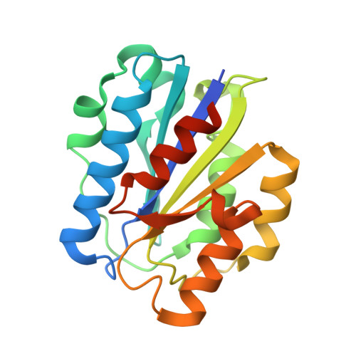
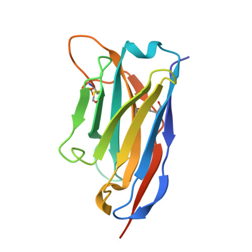
murine aM I-domain in complex with nanobody aCR3


Experimental Data Snapshot

  • Method: X-RAY DIFFRACTION
  • Resolution: 2.17 Å
  • R-Value Free: 
    0.259 (Depositor), 0.260 (DCC) 
  • R-Value Work: 
    0.202 (Depositor), 0.202 (DCC) 
  • R-Value Observed: 
    0.206 (Depositor) 

Starting Model: experimental
View more details

wwPDB Validation 3D Report Full Report

Validation slider image for 9HLL

Ligand Structure Quality Assessment 


This is version 1.1 of the entry. See complete history

Literature

Three cryo-EM structures of complement C3d-bound alpha M beta 2 reveal an unexpected layer of dynamics for alpha I-containing integrin receptors.

Lorentzen, J.Fruergaard, M.U.Lukacsi, S.Jorgensen, M.H.van Veghel, T.L.G.Jensen, R.K.Pietrzak-Lichwa, K.J.Bajtay, Z.Horejsi, V.Flygaard, R.K.Vorselen, D.Mortensen, S.A.Andersen, G.R.

(2026) Sci Adv 12: eaea7241-eaea7241

  • DOI: https://doi.org/10.1126/sciadv.aea7241
  • Primary Citation Related Structures: 
    9GMU, 9HLL, 9RM9, 9RMA, 9T3Y, 9T5V, 9T5W, 9T5Z

  • PubMed Abstract: 

    Integrins are heterodimeric membrane proteins acting as mechanosensing receptors. Nine human α-subunits contain a ligand binding αI domain, but how ligands activate αI integrins are not understood. We present cryo-EM structures of the αI integrin α M β 2 in complex with the C3d ligand. The ligand-bound αI domain appears to have two major opposite orientations relative to the β 2 subunit. Ligand binding induces an ordered conformation of the α M internal ligand region that is tightly packed between the α M β-propeller and the β 2 βI-domain. Recognition of the internal ligand induces an open βI conformation practically identical to that of ligand-bound αI-less integrins confirming that ligand binding and signaling are coupled by a universal mechanism across all integrins. Integration of our findings with prior data allows us to propose a model for C3dg/iC3b-bound α M β 2 in the phagocytotic cup and outline mechanistic models for external ligand-induced activation of α M β 2 .


  • Organizational Affiliation
    • Department of Molecular Biology and Genetics, Aarhus University, Aarhus, DK8000, Denmark.

Macromolecule Content 

  • Total Structure Weight: 65.98 kDa 
  • Atom Count: 4,407 
  • Modeled Residue Count: 550 
  • Deposited Residue Count: 588 
  • Unique protein chains: 2

Macromolecules

Find similar proteins by:|  3D Structure
Entity ID: 1
MoleculeChains  Sequence LengthOrganismDetailsImage
Integrin alpha-M198Mus musculusMutation(s): 0 
Gene Names: Itgam
UniProt & NIH Common Fund Data Resources
Find proteins for P05555 (Mus musculus)
Explore P05555 
Go to UniProtKB:  P05555
IMPC:  MGI:96607
Entity Groups
Sequence Clusters30% Identity50% Identity70% Identity90% Identity95% Identity100% Identity
UniProt GroupP05555
Sequence Annotations
Expand
Reference Sequence
Find similar proteins by:|  3D Structure
Entity ID: 2
MoleculeChains  Sequence LengthOrganismDetailsImage
aCR3 nanobody
B, C, D
130Lama glamaMutation(s): 0 

Experimental Data & Validation

Experimental Data

  • Method: X-RAY DIFFRACTION
  • Resolution: 2.17 Å
  • R-Value Free:  0.259 (Depositor), 0.260 (DCC) 
  • R-Value Work:  0.202 (Depositor), 0.202 (DCC) 
  • R-Value Observed: 0.206 (Depositor) 
Space Group: C 2 2 21
Unit Cell:
Length ( Å )Angle ( ˚ )
a = 78.402α = 90
b = 127.444β = 90
c = 125.855γ = 90
Software Package:
Software NamePurpose
PHENIXrefinement
XDSdata reduction
XSCALEdata scaling
PHASERphasing

Structure Validation

View Full Validation Report


Ligand Structure Quality Assessment 


Entry History 

& Funding Information

Deposition Data


Funding OrganizationLocationGrant Number
Other privateLeo LF-OC-22-001076
Other privateDenmarkAlexion Discovery RN7049
Other governmentDenmarkFNU 3103-00009B

Revision History  (Full details and data files)

  • Version 1.0: 2025-12-17
    Type: Initial release
  • Version 1.1: 2026-07-01
    Changes: Database references