9BJ2 | pdb_00009bj2

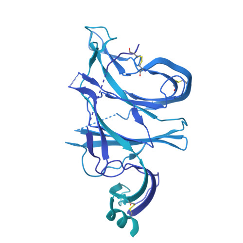
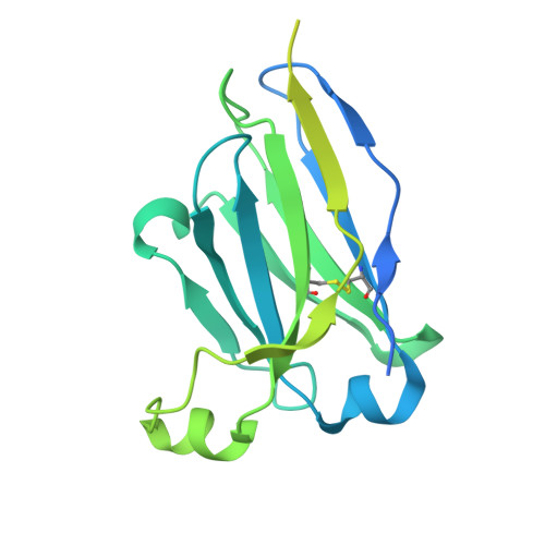
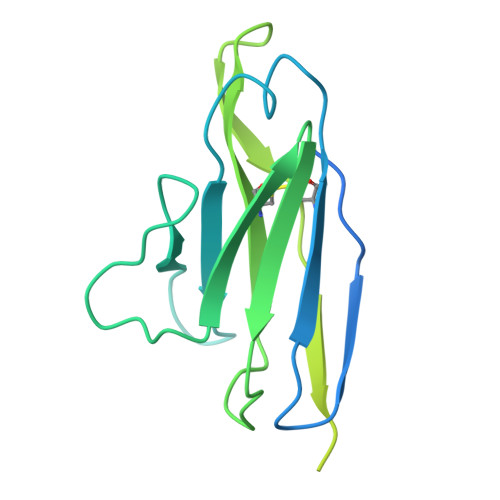
Structure of the SARS-CoV-2 S 6P trimer complex with the human neutralizing antibody Fab fragment, C1533 (local refinement of NTD and C1533)


Experimental Data Snapshot

  • Method: ELECTRON MICROSCOPY
  • Resolution: 2.80 Å
  • Aggregation State: PARTICLE 
  • Reconstruction Method: SINGLE PARTICLE 

wwPDB Validation 3D Report Full Report

Validation slider image for 9BJ2

This is version 1.1 of the entry. See complete history. 

Literature

Bispecific antibodies targeting the N-terminal and receptor binding domains potently neutralize SARS-CoV-2 variants of concern.

Rubio, A.A., Baharani, V.A., Dadonaite, B., Parada, M., Abernathy, M.E., Wang, Z., Lee, Y.E., Eso, M.R., Phung, J., Ramos, I., Chen, T., El Nesr, G., Bloom, J.D., Bieniasz, P.D., Nussenzweig, M.C., Barnes, C.O.

(2025) Sci Transl Med 17: eadq5720-eadq5720

  • DOI: https://doi.org/10.1126/scitranslmed.adq5720
  • Primary Citation Related Structures: 
    9BJ2, 9BJ3, 9BJ4

  • PubMed Abstract: 

    The ongoing emergence of severe acute respiratory syndrome coronavirus 2 (SARS-CoV-2) variants of concern (VOCs) that reduce the effectiveness of antibody therapeutics necessitates development of next-generation antibody modalities that are resilient to viral evolution. Here, we characterized amino-terminal domain (NTD)- and receptor binding domain (RBD)-specific monoclonal antibodies previously isolated from coronavirus disease 2019 (COVID-19) convalescent donors for their activity against emergent SARS-CoV-2 VOCs. Among these, the NTD-specific antibody C1596 displayed the greatest breadth of binding to VOCs, with cryo-electron microscopy structural analysis revealing recognition of a distinct NTD epitope outside of the site i antigenic supersite. Given C1596's favorable binding profile, we designed a series of bispecific antibodies (bsAbs), termed CoV2-biRNs, that featured both NTD and RBD specificities. Two of the C1596-inclusive bsAbs, CoV2-biRN5 and CoV2-biRN7, retained potent in vitro neutralization activity against all Omicron variants tested, including XBB.1.5, BA.2.86, and JN.1, contrasting the diminished potency of parental antibodies delivered as monotherapies or as a cocktail. Furthermore, prophylactic delivery of CoV2-biRN5 reduced the viral load within the lungs of K18-hACE2 mice after challenge with SARS-CoV-2 XBB.1.5. In conclusion, NTD-RBD bsAbs offer promising potential for the design of resilient, next-generation antibody therapeutics against SARS-CoV-2 VOCs.


  • Organizational Affiliation: 
    • Stanford Biosciences, Stanford School of Medicine, Stanford, CA 94305, USA.

Macromolecule Content 

  • Total Structure Weight: 195.77 kDa 
  • Atom Count: 3,920 
  • Modeled Residue Count: 501 
  • Deposited Residue Count: 1,784 
  • Unique protein chains: 3

Macromolecules

Find similar proteins by:|  3D Structure
Entity ID: 1
MoleculeChains  Sequence LengthOrganismDetailsImage
Spike glycoprotein1,288Severe acute respiratory syndrome coronavirus 2Mutation(s): 9 
Gene Names: S, 2
UniProt
Find proteins for P0DTC2 (Severe acute respiratory syndrome coronavirus 2)
Explore P0DTC2 
Go to UniProtKB:  P0DTC2
Entity Groups
Sequence Clusters30% Identity50% Identity70% Identity90% Identity95% Identity100% Identity
UniProt GroupP0DTC2
Glycosylation
Glycosylation Sites: 3Go to GlyGen: P0DTC2-1
Sequence Annotations
Expand
Reference Sequence
Find similar proteins by:|  3D Structure
Entity ID: 2
MoleculeChains  Sequence LengthOrganismDetailsImage
C1533 Heavy ChainB [auth H]261Homo sapiensMutation(s): 0 
Entity Groups
Sequence Clusters30% Identity50% Identity70% Identity90% Identity95% Identity100% Identity
Sequence Annotations
Expand
Reference Sequence
Find similar proteins by:|  3D Structure
Entity ID: 3
MoleculeChains  Sequence LengthOrganismDetailsImage
C1533 Light ChainC [auth L]235Homo sapiensMutation(s): 0 
Entity Groups
Sequence Clusters30% Identity50% Identity70% Identity90% Identity95% Identity100% Identity
Sequence Annotations
Expand
Reference Sequence

Experimental Data & Validation

Experimental Data

  • Method: ELECTRON MICROSCOPY
  • Resolution: 2.80 Å
  • Aggregation State: PARTICLE 
  • Reconstruction Method: SINGLE PARTICLE 
EM Software:
TaskSoftware PackageVersion
RECONSTRUCTIONcryoSPARC
MODEL REFINEMENTPHENIX

Structure Validation

View Full Validation Report


Entry History 

& Funding Information

Deposition Data


Funding OrganizationLocationGrant Number
Howard Hughes Medical Institute (HHMI)United StatesGT15335

Revision History  (Full details and data files)

  • Version 1.0: 2025-03-05
    Type: Initial release
  • Version 1.1: 2025-03-19
    Changes: Data collection, Database references