9XQR | pdb_00009xqr

Cryo-EM structure of P1X1 in complex with BTFA


Experimental Data Snapshot

  • Method: ELECTRON MICROSCOPY
  • Resolution: 3.14 Å
  • Aggregation State: PARTICLE 
  • Reconstruction Method: SINGLE PARTICLE 

wwPDB Validation 3D Report Full Report

Validation slider image for 9XQR

This is version 1.1 of the entry. See complete history

Literature

Structural basis for a central permeation pathway in the P2X1 receptor.

Zhang, H.Wu, P.Gu, Z.Xu, Y.Hu, W.Yuan, Q.Xia, B.Xu, H.E.Gao, Z.

(2026) Cell Discov 12

  • DOI: https://doi.org/10.1038/s41421-026-00881-w
  • Primary Citation Related Structures: 
    8ZT1, 8ZT2, 8ZT5, 8ZT8, 8ZTA, 8ZTD, 8ZTF, 9XQR

  • PubMed Abstract: 

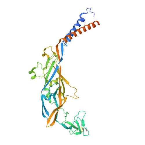
    The ion permeation pathway is a critical determinant of ion channel function and selectivity; however, the structural basis for ion permeation in the P2X1 receptor, an ATP-gated ion channel crucial for platelet activation, thrombosis, and male infertility, remains incompletely understood. Here, we present high-resolution cryo-electron microscopy (cryo-EM) structures of the P2X1 receptor, which reveal a central ion permeation pathway spanning the entire extracellular domain, complementing the existing paradigms of ion channel architecture for the P2X receptor family. Within this pathway, we identify specific sites that coordinate hydrated calcium ions, including an aspartate ring that acts as a selectivity filter at the apex of the central vestibule. We also discover that a small molecule, 3,5-bis(trifluoromethyl)aniline, binds at the top of the central vestibule and potently inhibits cation flux through this central permeation pathway. Our findings reveal a new inhibitor-binding site in the P2X1 receptor. These insights provide a structural framework for the rational design of subtype-specific P2X receptor inhibitors targeting the central vestibule.


  • Organizational Affiliation
    • The State Key Laboratory of Drug Research, Shanghai Institute of Materia Medica, Chinese Academy of Sciences, Shanghai, China.

Macromolecule Content 

  • Total Structure Weight: 147.77 kDa 
  • Atom Count: 7,995 
  • Modeled Residue Count: 997 
  • Deposited Residue Count: 1,281 
  • Unique protein chains: 1

Macromolecules

Find similar proteins by:|  3D Structure
Entity ID: 1
MoleculeChains  Sequence LengthOrganismDetailsImage
P2X purinoceptor 1A [auth B],
B [auth C],
C [auth A]
427Mus musculusMutation(s): 0 
Gene Names: P2rx1
UniProt
Find proteins for P51576 (Mus musculus)
Explore P51576 
Go to UniProtKB:  P51576
Entity Groups
Sequence Clusters30% Identity50% Identity70% Identity90% Identity95% Identity100% Identity
UniProt GroupP51576
Sequence Annotations
Expand
Reference Sequence

Small Molecules

Ligands 5 Unique
IDChains Name / Formula / InChI Key2D Diagram3D Interactions
ATP

Query on ATP


Download:Ideal Coordinates CCD File
D [auth B],
F [auth B],
J [auth A]
ADENOSINE-5'-TRIPHOSPHATE
C10 H16 N5 O13 P3
ZKHQWZAMYRWXGA-KQYNXXCUSA-N
A1D8Z
(Subject of Investigation/LOI)

Query on A1D8Z


Download:Ideal Coordinates CCD File
M [auth A]3,5-bis(trifluoromethyl)aniline
C8 H5 F6 N
CDIDGWDGQGVCIB-UHFFFAOYSA-N
NAG

Query on NAG


Download:Ideal Coordinates CCD File
E [auth B],
K [auth A],
L [auth A]
2-acetamido-2-deoxy-beta-D-glucopyranose
C8 H15 N O6
OVRNDRQMDRJTHS-FMDGEEDCSA-N
CA

Query on CA


Download:Ideal Coordinates CCD File
G [auth B]CALCIUM ION
Ca
BHPQYMZQTOCNFJ-UHFFFAOYSA-N
MG

Query on MG


Download:Ideal Coordinates CCD File
H [auth B],
I [auth C],
N [auth A]
MAGNESIUM ION
Mg
JLVVSXFLKOJNIY-UHFFFAOYSA-N

Experimental Data & Validation

Experimental Data

  • Method: ELECTRON MICROSCOPY
  • Resolution: 3.14 Å
  • Aggregation State: PARTICLE 
  • Reconstruction Method: SINGLE PARTICLE 
EM Software:
TaskSoftware PackageVersion
RECONSTRUCTIONcryoSPARC

Structure Validation

View Full Validation Report


Entry History 

& Funding Information

Deposition Data

  • Released Date: 2025-12-24 
  • Deposition Author(s): Zhang, H.

Funding OrganizationLocationGrant Number
National Natural Science Foundation of China (NSFC)China--

Revision History  (Full details and data files)

  • Version 1.0: 2025-12-24
    Type: Initial release
  • Version 1.1: 2026-06-24
    Changes: Data collection, Database references