9I5A | pdb_00009i5a

Crystal structure of wild type perlecan region 3 construct I876-V1272 construct including one laminin IV-like and four laminin EGF-like domains.


Experimental Data Snapshot

  • Method: X-RAY DIFFRACTION
  • Resolution: 2.04 Å
  • R-Value Free: 
    0.209 (Depositor), 0.209 (DCC) 
  • R-Value Work: 
    0.178 (Depositor), 0.178 (DCC) 

Starting Model: in silico
View more details

wwPDB Validation   3D Report Full Report


Ligand Structure Quality Assessment 


This is version 1.1 of the entry. See complete history


Literature

Structural insights on perlecan and Schwartz-Jampel syndrome.

Sohail, A.A.Koski, M.K.Ruddock, L.W.

(2025) Matrix Biol 138: 1-7

  • DOI: https://doi.org/10.1016/j.matbio.2025.03.002
  • Primary Citation of Related Structures:  
    9I5A, 9I5B

  • PubMed Abstract: 

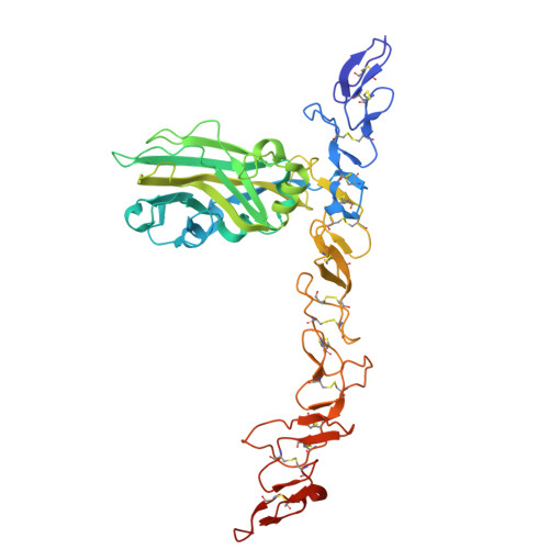
    Perlecan is an essential multi-domain, disulfide bond rich basement membrane protein. Mutations in perlecan cause Schwartz-Jampel syndrome and dyssegmental dysplasia. While there has been a large body of experimental work reported on perlecan, there is only minimal structural information available to date. There is no prior structural data for region 3 of perlecan in which some Schwartz-Jampel syndrome causing point mutations have been reported. Here, we produce constructs of the disulfide rich region 3 of perlecan along with five mutations previously reported to cause Schwatz-Jampel syndrome. Four of the mutations resulted in decreased yields and thermal stability compared to the wild-type protein. In contrast, the P1019L mutation was produced in good yields and showed higher thermal stability than the wild-type protein. The crystal structures for both the wild-type and P1019L mutation were solved. As expected, both showed laminin IV-like and laminin-type EGF-like domains, with the P1019L mutation resulting in only a minor conformational change in a loop region and no significant changes in regular secondary or tertiary structure.


  • Organizational Affiliation
    • Faculty of Biochemistry and Molecular Medicine, University of Oulu, Oulu, 90220, Finland.

Macromolecules
Find similar proteins by:  (by identity cutoff)  |  3D Structure
Entity ID: 1
MoleculeChains Sequence LengthOrganismDetailsImage
Basement membrane-specific heparan sulfate proteoglycan core protein
A, B
397Mus musculusMutation(s): 0 
Gene Names: Hspg2
UniProt
Find proteins for Q05793 (Mus musculus)
Explore Q05793 
Go to UniProtKB:  Q05793
Entity Groups  
Sequence Clusters30% Identity50% Identity70% Identity90% Identity95% Identity100% Identity
UniProt GroupQ05793
Sequence Annotations
Expand
  • Reference Sequence
Small Molecules
Ligands 2 Unique
IDChains Name / Formula / InChI Key2D Diagram3D Interactions
SIN (Subject of Investigation/LOI)
Query on SIN

Download Ideal Coordinates CCD File 
C [auth A],
G [auth B]
SUCCINIC ACID
C4 H6 O4
KDYFGRWQOYBRFD-UHFFFAOYSA-N
GOL (Subject of Investigation/LOI)
Query on GOL

Download Ideal Coordinates CCD File 
D [auth A]
E [auth A]
F [auth A]
H [auth B]
I [auth B]
D [auth A],
E [auth A],
F [auth A],
H [auth B],
I [auth B],
J [auth B],
K [auth B]
GLYCEROL
C3 H8 O3
PEDCQBHIVMGVHV-UHFFFAOYSA-N
Experimental Data & Validation

Experimental Data

  • Method: X-RAY DIFFRACTION
  • Resolution: 2.04 Å
  • R-Value Free:  0.209 (Depositor), 0.209 (DCC) 
  • R-Value Work:  0.178 (Depositor), 0.178 (DCC) 
Space Group: P 1
Unit Cell:
Length ( Å )Angle ( ˚ )
a = 57.637α = 74.384
b = 74.612β = 74.084
c = 78.769γ = 79.729
Software Package:
Software NamePurpose
REFMACrefinement
XDSdata reduction
XDSdata scaling
PHENIXphasing

Structure Validation

View Full Validation Report


Ligand Structure Quality Assessment 


Entry History & Funding Information

Deposition Data


Funding OrganizationLocationGrant Number
Academy of FinlandFinland272573

Revision History  (Full details and data files)

  • Version 1.0: 2025-04-02
    Type: Initial release
  • Version 1.1: 2025-05-21
    Changes: Database references