9H6X | pdb_00009h6x

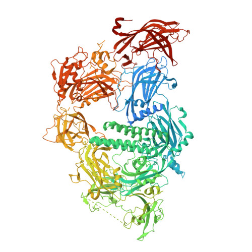
Cryo-EM structure of lipid-bound human myoferlin (25 mol% DOPS/5 mol% PI(4,5)P2 nanodisc)


Experimental Data Snapshot

  • Method: ELECTRON MICROSCOPY
  • Resolution: 2.56 Å
  • Aggregation State: PARTICLE 
  • Reconstruction Method: SINGLE PARTICLE 

Starting Model: in silico
View more details

wwPDB Validation   3D Report Full Report


This is version 1.0 of the entry. See complete history


Literature

Structural insights into lipid membrane binding by human ferlins.

Cretu, C.Chernev, A.Kibedi Szabo, C.Z.Pena, V.Urlaub, H.Moser, T.Preobraschenski, J.

(2025) EMBO J 

  • DOI: https://doi.org/10.1038/s44318-025-00463-8
  • Primary Citation of Related Structures:  
    9H6X, 9QKV, 9QLE, 9QLF, 9QLN, 9QLS

  • PubMed Abstract: 

    Ferlins are ancient membrane proteins with a unique architecture, and play central roles in crucial processes that involve Ca 2+ -dependent vesicle fusion. Despite their links to multiple human diseases and numerous functional studies, a mechanistic understanding of how these multi-C 2 domain-containing proteins interact with lipid membranes to promote membrane remodelling and fusion is currently lacking. Here we obtain near-complete cryo-electron microscopy structures of human myoferlin and dysferlin in their Ca 2+ - and lipid-bound states. We show that ferlins adopt compact, ring-like tertiary structures upon membrane binding. The top arch of the ferlin ring, composed of the C 2 C-C 2 D region, is rigid and exhibits only little variability across the observed functional states. In contrast, the N-terminal C 2 B and the C-terminal C 2 F-C 2 G domains cycle between alternative conformations and, in response to Ca 2+ , close the ferlin ring, promoting tight interaction with the target membrane. Probing key domain interfaces validates the observed architecture, and informs a model of how ferlins engage lipid bilayers in a Ca 2+ -dependent manner. This work reveals the general principles of human ferlin structures and provides a framework for future analyses of ferlin-dependent cellular functions and disease mechanisms.


  • Organizational Affiliation

    Institute for Auditory Neuroscience and InnerEarLab, University Medical Center Göttingen, Göttingen, Germany. constantin.cretu@med.uni-goettingen.de.


Macromolecules
Find similar proteins by:  (by identity cutoff)  |  3D Structure
Entity ID: 1
MoleculeChains Sequence LengthOrganismDetailsImage
MyoferlinA [auth D]2,048Homo sapiensMutation(s): 0 
Gene Names: MYOFFER1L3KIAA1207
UniProt & NIH Common Fund Data Resources
Find proteins for Q9NZM1 (Homo sapiens)
Explore Q9NZM1 
Go to UniProtKB:  Q9NZM1
PHAROS:  Q9NZM1
GTEx:  ENSG00000138119 
Entity Groups  
Sequence Clusters30% Identity50% Identity70% Identity90% Identity95% Identity100% Identity
UniProt GroupQ9NZM1
Sequence Annotations
Expand
  • Reference Sequence
Small Molecules
Ligands 2 Unique
IDChains Name / Formula / InChI Key2D Diagram3D Interactions
PSF (Subject of Investigation/LOI)
Query on PSF

Download Ideal Coordinates CCD File 
B [auth D],
M [auth D]
1,2-DICAPROYL-SN-PHOSPHATIDYL-L-SERINE
C18 H34 N O10 P
MIQYPPGTNIFAPO-CABCVRRESA-N
CA (Subject of Investigation/LOI)
Query on CA

Download Ideal Coordinates CCD File 
C [auth D]
D
E [auth D]
F [auth D]
G [auth D]
C [auth D],
D,
E [auth D],
F [auth D],
G [auth D],
H [auth D],
I [auth D],
J [auth D],
K [auth D],
L [auth D]
CALCIUM ION
Ca
BHPQYMZQTOCNFJ-UHFFFAOYSA-N
Experimental Data & Validation

Experimental Data

  • Method: ELECTRON MICROSCOPY
  • Resolution: 2.56 Å
  • Aggregation State: PARTICLE 
  • Reconstruction Method: SINGLE PARTICLE 
EM Software:
TaskSoftware PackageVersion
RECONSTRUCTIONcryoSPARCv4.5
MODEL REFINEMENTPHENIX1.21.2_5419

Structure Validation

View Full Validation Report


Entry History & Funding Information

Deposition Data


Funding OrganizationLocationGrant Number
German Research Foundation (DFG)GermanyCR 937/2-1, EXC 2067/1-390729940

Revision History  (Full details and data files)

  • Version 1.0: 2025-06-04
    Type: Initial release