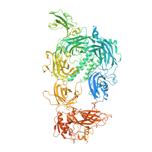
Structural insights into lipid membrane binding by human ferlins.
Cretu, C., Chernev, A., Kibedi Szabo, C.Z., Pena, V., Urlaub, H., Moser, T., Preobraschenski, J.(2025) EMBO J 44: 3926-3958
- PubMed: 40437073 Search on PubMedSearch on PubMed Central
- DOI: https://doi.org/10.1038/s44318-025-00463-8
- Primary Citation Related Structures:
9H6X, 9QKV, 9QLE, 9QLF, 9QLN, 9QLS - PubMed Abstract:
Ferlins are ancient membrane proteins with a unique architecture, and play central roles in crucial processes that involve Ca 2+ -dependent vesicle fusion. Despite their links to multiple human diseases and numerous functional studies, a mechanistic understanding of how these multi-C 2 domain-containing proteins interact with lipid membranes to promote membrane remodelling and fusion is currently lacking. Here we obtain near-complete cryo-electron microscopy structures of human myoferlin and dysferlin in their Ca 2+ - and lipid-bound states. We show that ferlins adopt compact, ring-like tertiary structures upon membrane binding. The top arch of the ferlin ring, composed of the C 2 C-C 2 D region, is rigid and exhibits only little variability across the observed functional states. In contrast, the N-terminal C 2 B and the C-terminal C 2 F-C 2 G domains cycle between alternative conformations and, in response to Ca 2+ , close the ferlin ring, promoting tight interaction with the target membrane. Probing key domain interfaces validates the observed architecture, and informs a model of how ferlins engage lipid bilayers in a Ca 2+ -dependent manner. This work reveals the general principles of human ferlin structures and provides a framework for future analyses of ferlin-dependent cellular functions and disease mechanisms.
- Institute for Auditory Neuroscience and InnerEarLab, University Medical Center Göttingen, Göttingen, Germany. constantin.cretu@med.uni-goettingen.de.
Organizational Affiliation: