9EV0 | pdb_00009ev0

Structure of the AAP filament of Sulfolobus acidocaldarius strain MW039 (delta agl3 mutant).


Experimental Data Snapshot

  • Method: ELECTRON MICROSCOPY
  • Resolution: 2.38 Å
  • Aggregation State: FILAMENT 
  • Reconstruction Method: HELICAL 

wwPDB Validation   3D Report Full Report


This is version 1.0 of the entry. See complete history


Literature

Towards a molecular picture of the archaeal cell surface.

Gaines, M.C.Isupov, M.N.McLaren, M.Mollat, C.L.Haque, R.U.Stephenson, J.K.Sivabalasarma, S.Hanus, C.Kattnig, D.Gold, V.A.M.Albers, S.Daum, B.

(2024) Nat Commun 15: 10401-10401

  • DOI: https://doi.org/10.1038/s41467-024-53986-9
  • Primary Citation of Related Structures:  
    8QX4, 8RZL, 9ETS, 9ETT, 9EV0

  • PubMed Abstract: 

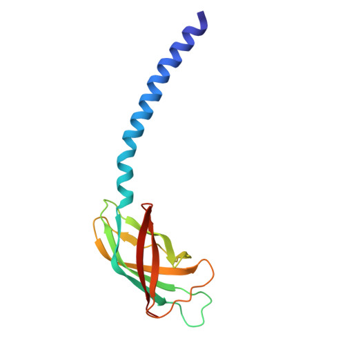
    Archaea produce various protein filaments with specialised functions. While some archaea produce only one type of filament, the archaeal model species Sulfolobus acidocaldarius generates four. These include rotary swimming propellers analogous to bacterial flagella (archaella), pili for twitching motility (Aap), adhesive fibres (threads), and filaments facilitating homologous recombination upon UV stress (UV pili). Here, we use cryo-electron microscopy to describe the structure of the S. acidocaldarius archaellum at 2.0 Å resolution, and update the structures of the thread and the Aap pilus at 2.7 Å and 2.6 Å resolution, respectively. We define features unique to archaella of the order Sulfolobales and compare their structure to those of Aap and threads in the context of the S-layer. We define distinct N-glycan patterns in the three filaments and identify a putative O-glycosylation site in the thread. Finally, we ascertain whether N-glycan truncation leads to structural changes in archaella and Aap.


  • Organizational Affiliation
    • Living Systems Institute, University of Exeter, Exeter, UK.

Macromolecules
Find similar proteins by:  (by identity cutoff)  |  3D Structure
Entity ID: 1
MoleculeChains Sequence LengthOrganismDetailsImage
DUF4352 domain-containing protein141Sulfolobus acidocaldariusMutation(s): 0 
UniProt
Find proteins for Q4J6H7 (Sulfolobus acidocaldarius (strain ATCC 33909 / DSM 639 / JCM 8929 / NBRC 15157 / NCIMB 11770))
Explore Q4J6H7 
Go to UniProtKB:  Q4J6H7
Entity Groups  
Sequence Clusters30% Identity50% Identity70% Identity90% Identity95% Identity100% Identity
UniProt GroupQ4J6H7
Glycosylation
Glycosylation Sites: 3
Sequence Annotations
Expand
  • Reference Sequence
Oligosaccharides

Help

Entity ID: 2
MoleculeChains Length2D Diagram Glycosylation3D Interactions
alpha-D-mannopyranose-(1-6)-2-acetamido-2-deoxy-beta-D-glucopyranose-(1-4)-2-acetamido-2-deoxy-beta-D-glucopyranose
AB [auth 0],
AC [auth QA],
AD [auth qA],
BB [auth 1],
BC [auth RA],
AB [auth 0],
AC [auth QA],
AD [auth qA],
BB [auth 1],
BC [auth RA],
BD [auth rA],
CB [auth 2],
CC [auth SA],
CD [auth sA],
DB [auth 3],
DC [auth TA],
DD [auth tA],
EA [auth e],
EB [auth 4],
EC [auth UA],
ED [auth uA],
FA [auth f],
FB [auth 5],
FC [auth VA],
FD [auth vA],
GA [auth g],
GB [auth 6],
GC [auth WA],
GD [auth wA],
HA [auth h],
HB [auth 7],
HC [auth XA],
HD [auth xA],
IA [auth i],
IB [auth 8],
IC [auth YA],
ID [auth yA],
JA [auth j],
JB [auth 9],
JC [auth ZA],
JD [auth zA],
KA [auth k],
KB [auth AA],
KC [auth aA],
KD [auth 0A],
LA [auth l],
LB [auth BA],
LC [auth bA],
LD [auth 1A],
MA [auth m],
MB [auth CA],
MC [auth cA],
MD [auth 2A],
NA [auth n],
NB [auth DA],
NC [auth dA],
ND [auth 3A],
OA [auth o],
OB [auth EA],
OC [auth eA],
OD [auth 4A],
PA [auth p],
PB [auth FA],
PC [auth fA],
PD [auth 5A],
QA [auth q],
QB [auth GA],
QC [auth gA],
RA [auth r],
RB [auth HA],
RC [auth hA],
SA [auth s],
SB [auth IA],
SC [auth iA],
TA [auth t],
TB [auth JA],
TC [auth jA],
UA [auth u],
UB [auth KA],
UC [auth kA],
VA [auth v],
VB [auth LA],
VC [auth lA],
WA [auth w],
WB [auth MA],
WC [auth mA],
XA [auth x],
XB [auth NA],
XC [auth nA],
YA [auth y],
YB [auth OA],
YC [auth oA],
ZA [auth z],
ZB [auth PA],
ZC [auth pA]
3N-Glycosylation
Glycosylation Resources
GlyTouCan:  G34031ES
GlyCosmos:  G34031ES
GlyGen:  G34031ES
Experimental Data & Validation

Experimental Data

  • Method: ELECTRON MICROSCOPY
  • Resolution: 2.38 Å
  • Aggregation State: FILAMENT 
  • Reconstruction Method: HELICAL 
EM Software:
TaskSoftware PackageVersion
MODEL REFINEMENTREFMAC5.8.0267

Structure Validation

View Full Validation Report


Entry History & Funding Information

Deposition Data


Funding OrganizationLocationGrant Number
European Research Council (ERC)European Union109784

Revision History  (Full details and data files)

  • Version 1.0: 2025-02-05
    Type: Initial release