9AVG | pdb_00009avg

Structure of human calcium-sensing receptor in complex with chimeric Gs (miniGis) protein in nanodiscs


Experimental Data Snapshot

  • Method: ELECTRON MICROSCOPY
  • Resolution: 3.60 Å
  • Aggregation State: PARTICLE 
  • Reconstruction Method: SINGLE PARTICLE 

Starting Models: experimental
View more details

wwPDB Validation 3D Report Full Report

Validation slider image for 9AVG

This is version 1.3 of the entry. See complete history

Literature

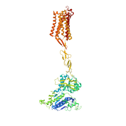
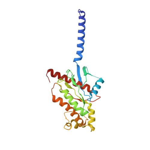
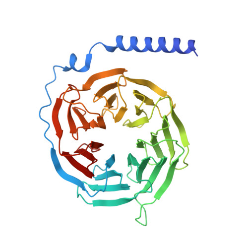
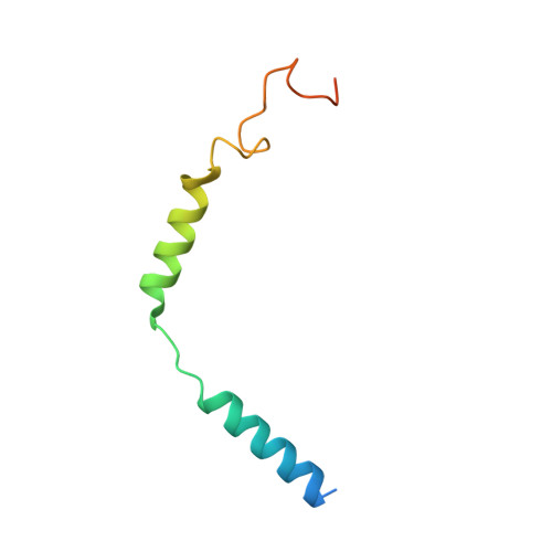
Promiscuous G-protein activation by the calcium-sensing receptor.

Zuo, H.Park, J.Frangaj, A.Ye, J.Lu, G.Manning, J.J.Asher, W.B.Lu, Z.Hu, G.B.Wang, L.Mendez, J.Eng, E.Zhang, Z.Lin, X.Grassucci, R.Hendrickson, W.A.Clarke, O.B.Javitch, J.A.Conigrave, A.D.Fan, Q.R.

(2024) Nature 629: 481-488

  • DOI: https://doi.org/10.1038/s41586-024-07331-1
  • Primary Citation Related Structures: 
    9ASB, 9AVG, 9AVL, 9AXF, 9AYF

  • PubMed Abstract: 

    The human calcium-sensing receptor (CaSR) detects fluctuations in the extracellular Ca 2+ concentration and maintains Ca 2+ homeostasis 1,2 . It also mediates diverse cellular processes not associated with Ca 2+ balance 3-5 . The functional pleiotropy of CaSR arises in part from its ability to signal through several G-protein subtypes 6 . We determined structures of CaSR in complex with G proteins from three different subfamilies: G q , G i and G s . We found that the homodimeric CaSR of each complex couples to a single G protein through a common mode. This involves the C-terminal helix of each Gα subunit binding to a shallow pocket that is formed in one CaSR subunit by all three intracellular loops (ICL1-ICL3), an extended transmembrane helix 3 and an ordered C-terminal region. G-protein binding expands the transmembrane dimer interface, which is further stabilized by phospholipid. The restraint imposed by the receptor dimer, in combination with ICL2, enables G-protein activation by facilitating conformational transition of Gα. We identified a single Gα residue that determines G q and G s versus G i selectivity. The length and flexibility of ICL2 allows CaSR to bind all three Gα subtypes, thereby conferring capacity for promiscuous G-protein coupling.


  • Organizational Affiliation
    • Department of Molecular Pharmacology and Therapeutics, Columbia University, New York, NY, USA.

Macromolecule Content 

  • Total Structure Weight: 285.94 kDa 
  • Atom Count: 17,498 
  • Modeled Residue Count: 2,208 
  • Deposited Residue Count: 2,487 
  • Unique protein chains: 4

Macromolecules

Find similar proteins by:|  3D Structure
Entity ID: 1
MoleculeChains  Sequence LengthOrganismDetailsImage
Isoform 1 of Extracellular calcium-sensing receptorA [auth Q],
B [auth R]
911Homo sapiensMutation(s): 0 
Gene Names: CASRGPRC2APCAR1
Membrane Entity: Yes 
UniProt & NIH Common Fund Data Resources
Find proteins for P41180 (Homo sapiens)
Explore P41180 
Go to UniProtKB:  P41180
PHAROS:  P41180
GTEx:  ENSG00000036828 
Entity Groups
Sequence Clusters30% Identity50% Identity70% Identity90% Identity95% Identity100% Identity
UniProt GroupP41180
Glycosylation
Glycosylation Sites: 4Go to GlyGen: P41180-1
Sequence Annotations
Expand
Reference Sequence
Find similar proteins by:|  3D Structure
Entity ID: 2
MoleculeChains  Sequence LengthOrganismDetailsImage
Chimeric mini guanine nucleotide-binding protein G(i)(s) subunit alphaC [auth A]246Homo sapiensMutation(s): 21 
Gene Names: GNAI1GNAS
EC: 3.6.5
Membrane Entity: Yes 
UniProt & NIH Common Fund Data Resources
Find proteins for P63092 (Homo sapiens)
Explore P63092 
Go to UniProtKB:  P63092
PHAROS:  P63092
GTEx:  ENSG00000087460 
Find proteins for P63096 (Homo sapiens)
Explore P63096 
Go to UniProtKB:  P63096
PHAROS:  P63096
GTEx:  ENSG00000127955 
Entity Groups
Sequence Clusters30% Identity50% Identity70% Identity90% Identity95% Identity100% Identity
UniProt GroupsP63096P63092
Sequence Annotations
Expand
Reference Sequence
Find similar proteins by:|  3D Structure
Entity ID: 3
MoleculeChains  Sequence LengthOrganismDetailsImage
Guanine nucleotide-binding protein G(I)/G(S)/G(T) subunit beta-1D [auth B]348Homo sapiensMutation(s): 0 
Gene Names: GNB1
UniProt & NIH Common Fund Data Resources
Find proteins for P62873 (Homo sapiens)
Explore P62873 
Go to UniProtKB:  P62873
PHAROS:  P62873
GTEx:  ENSG00000078369 
Entity Groups
Sequence Clusters30% Identity50% Identity70% Identity90% Identity95% Identity100% Identity
UniProt GroupP62873
Sequence Annotations
Expand
Reference Sequence
Find similar proteins by:|  3D Structure
Entity ID: 4
MoleculeChains  Sequence LengthOrganismDetailsImage
Guanine nucleotide-binding protein G(I)/G(S)/G(O) subunit gamma-2E [auth G]71Homo sapiensMutation(s): 0 
Gene Names: GNG2
Membrane Entity: Yes 
UniProt & NIH Common Fund Data Resources
Find proteins for P59768 (Homo sapiens)
Explore P59768 
Go to UniProtKB:  P59768
PHAROS:  P59768
GTEx:  ENSG00000186469 
Entity Groups
Sequence Clusters30% Identity50% Identity70% Identity90% Identity95% Identity100% Identity
UniProt GroupP59768
Sequence Annotations
Expand
Reference Sequence

Oligosaccharides

Help  
Entity ID: 5
MoleculeChains Length2D Diagram GlycosylationD Interactions
2-acetamido-2-deoxy-beta-D-glucopyranose-(1-4)-2-acetamido-2-deoxy-beta-D-glucopyranoseF [auth C],
G [auth D],
H [auth E],
I [auth F]
2N-Glycosylation
Glycosylation Resources
GlyTouCan: G42666HT
GlyCosmos: G42666HT
GlyGen: G42666HT

Small Molecules

Ligands 7 Unique
IDChains Name / Formula / InChI Key2D Diagram3D Interactions
A1AF7

Query on A1AF7


Download:Ideal Coordinates CCD File
BA [auth R](19R,22S,25R)-22,25,26-trihydroxy-16,22-dioxo-17,21,23-trioxa-22lambda~5~-phosphahexacosan-19-yl (9Z)-octadec-9-enoate
C40 H77 O10 P
PAZGBAOHGQRCBP-LLSCNKDYSA-N
Y01

Query on Y01


Download:Ideal Coordinates CCD File
S [auth Q]CHOLESTEROL HEMISUCCINATE
C31 H50 O4
WLNARFZDISHUGS-MIXBDBMTSA-N
9IG

Query on 9IG


Download:Ideal Coordinates CCD File
AA [auth R],
R [auth Q]
3-(2-chlorophenyl)-N-[(1R)-1-(3-methoxyphenyl)ethyl]propan-1-amine
C18 H22 Cl N O
ZVQUCWXZCKWZBP-CQSZACIVSA-N
NAG

Query on NAG


Download:Ideal Coordinates CCD File
J [auth Q],
K [auth Q],
L [auth Q],
T [auth R],
U [auth R]
2-acetamido-2-deoxy-beta-D-glucopyranose
C8 H15 N O6
OVRNDRQMDRJTHS-FMDGEEDCSA-N
TCR

Query on TCR


Download:Ideal Coordinates CCD File
M [auth Q],
V [auth R]
CYCLOMETHYLTRYPTOPHAN
C12 H12 N2 O2
FSNCEEGOMTYXKY-JTQLQIEISA-N
PO4

Query on PO4


Download:Ideal Coordinates CCD File
N [auth Q],
W [auth R]
PHOSPHATE ION
O4 P
NBIIXXVUZAFLBC-UHFFFAOYSA-K
CA

Query on CA


Download:Ideal Coordinates CCD File
O [auth Q]
P [auth Q]
Q
X [auth R]
Y [auth R]
O [auth Q],
P [auth Q],
Q,
X [auth R],
Y [auth R],
Z [auth R]
CALCIUM ION
Ca
BHPQYMZQTOCNFJ-UHFFFAOYSA-N
Binding Affinity Annotations 
IDSourceBinding Affinity
9IG BindingDB:  9AVG EC50: min: 80, max: 194 (nM) from 2 assay(s)

Experimental Data & Validation

Experimental Data

  • Method: ELECTRON MICROSCOPY
  • Resolution: 3.60 Å
  • Aggregation State: PARTICLE 
  • Reconstruction Method: SINGLE PARTICLE 
EM Software:
TaskSoftware PackageVersion
RECONSTRUCTIONcryoSPARC3.3.2
MODEL REFINEMENTPHENIX1.19.2_4158:

Structure Validation

View Full Validation Report


Entry History 

& Funding Information

Deposition Data


Funding OrganizationLocationGrant Number
National Institutes of Health/National Institute of General Medical Sciences (NIH/NIGMS)United StatesR35GM141871

Revision History  (Full details and data files)

  • Version 1.0: 2024-04-17
    Type: Initial release
  • Version 1.1: 2024-05-01
    Changes: Database references
  • Version 1.2: 2024-05-22
    Changes: Database references
  • Version 1.3: 2024-10-30
    Changes: Data collection, Structure summary